Anatomically Consistent TMJ Disc Segmentation via Semantic Anchoring and Clinical Priors
Publication
Metrics
Paper Preview
Abstract
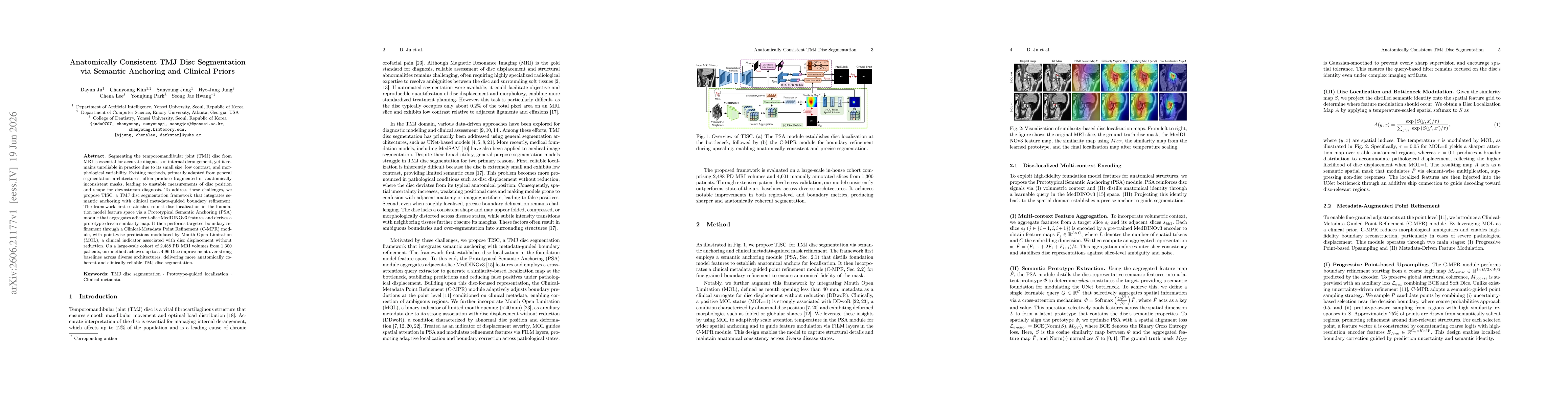
Segmenting the temporomandibular joint (TMJ) disc from MRI is essential for accurate diagnosis of internal derangement, yet it remains unreliable in practice due to its small size, low contrast, and morphological variability. Existing methods, primarily adapted from general segmentation architectures, often produce fragmented or anatomically inconsistent masks, leading to unstable measurements of disc position and shape for downstream diagnosis. To address these challenges, we propose TISC, a TMJ disc segmentation framework that integrates semantic anchoring with clinical metadata-guided boundary refinement. The framework first establishes robust disc localization in the foundation model feature space via a Prototypical Semantic Anchoring (PSA) module that aggregates adjacent-slice MedDINOv3 features and derives a prototype-driven similarity map. It then performs targeted boundary refinement through a Clinical-Metadata Point Refinement (C-MPR) module, with point-wise predictions modulated by Mouth Open Limitation (MOL), a clinical indicator associated with disc displacement without reduction. On a large-scale cohort of 2,488 PD MRI volumes from 1,300 patients, our method achieves up to a 4.96 Dice improvement over strong baselines across diverse architectures, delivering more anatomically coherent and clinically reliable TMJ disc segmentation.
AI Key Findings
Get AI-generated insights about this paper's methodology, results, significance, and more — seven facets brought into focus.
Discussion 0