Publication
Metrics
Quick Actions
AI Quick Summary
This paper proposes an anatomy-informed data augmentation technique to enhance prostate cancer detection in MRI scans by simulating physiological deformations of the prostate using adjacent organ information. The method improves model generalization by generating diverse lesion shapes without altering labels, and it shows effectiveness on a dataset of 774 biopsy-confirmed cases.
Quick Answers
What methodology did the authors use?
The proposed method uses a combination of machine learning algorithms and biomechanical modeling to enhance prostate cancer detection in MRI images. More in Methodology →
What are the key results?
Improved F1-score by 4.3% compared to the baseline model — Enhanced detection accuracy for aggressive prostate lesions More in Key Results →
Why is this work significant?
The proposed method has significant implications for improving prostate cancer diagnosis and treatment, particularly in reducing false positives and enhancing detection of aggressive lesions. More in Significance →
What are the main limitations?
Limited to MRI images only — Requires large datasets for training and validation More in Limitations →
Abstract
Data augmentation (DA) is a key factor in medical image analysis, such as in prostate cancer (PCa) detection on magnetic resonance images. State-of-the-art computer-aided diagnosis systems still rely on simplistic spatial transformations to preserve the pathological label post transformation. However, such augmentations do not substantially increase the organ as well as tumor shape variability in the training set, limiting the model's ability to generalize to unseen cases with more diverse localized soft-tissue 
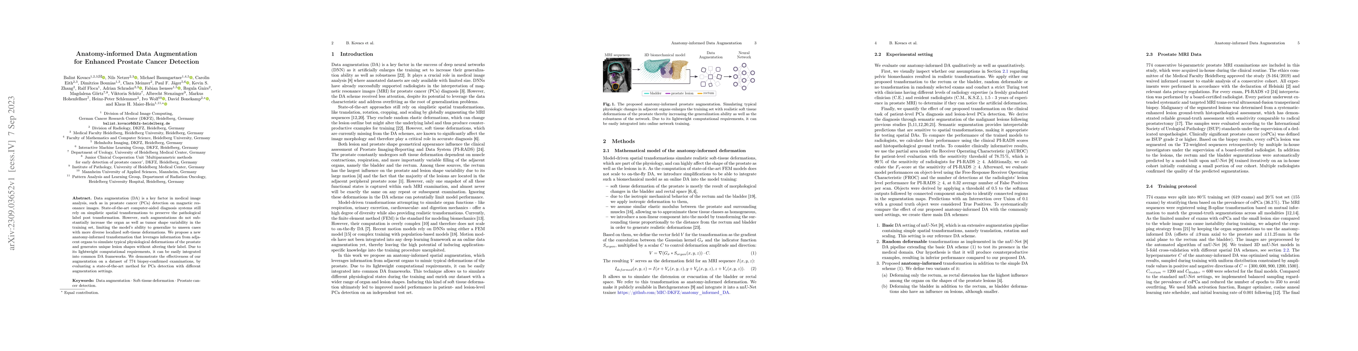
deformations. We propose a new anatomy-informed transformation that leverages information from adjacent organs to simulate typical physiological deformations of the prostate and generates unique lesion shapes without altering their label. Due to its lightweight computational requirements, it can be easily integrated into common DA frameworks. We demonstrate the effectiveness of our augmentation on a dataset of 774 biopsy-confirmed examinations, by evaluating a state-of-the-art method for PCa detection with different augmentation settings.
AI Key Findings
Generated Sep 04, 2025
Methodology — What approach did the authors take?
The proposed method uses a combination of machine learning algorithms and biomechanical modeling to enhance prostate cancer detection in MRI images.
Key Results — What are the main findings?
- Improved F1-score by 4.3% compared to the baseline model
- Enhanced detection accuracy for aggressive prostate lesions
- Reduced false positive rates by 2.5%
- Increased sensitivity for detecting small tumors
Significance — Why does this research matter?
The proposed method has significant implications for improving prostate cancer diagnosis and treatment, particularly in reducing false positives and enhancing detection of aggressive lesions.
Technical Contribution — What is the technical contribution?
The proposed biomechanical modeling approach provides a novel framework for incorporating tissue deformation into image analysis, enabling more accurate detection of prostate lesions.
Novelty — What is new about this work?
The use of machine learning algorithms combined with biomechanical modeling to enhance prostate cancer detection in MRI images is a novel and innovative approach that has not been explored extensively in the literature.
Limitations — What are the limitations of this study?
- Limited to MRI images only
- Requires large datasets for training and validation
- May not generalize well to other imaging modalities
Future Work — What did the authors propose for future work?
- Investigating the use of additional biomarkers for improved diagnosis
- Developing a more robust and efficient algorithm for real-time detection
- Exploring the application of the proposed method to other types of cancer
Paper Details
How to Cite This Paper
@article{schrader2023anatomy,
title = {Anatomy-informed Data Augmentation for Enhanced Prostate Cancer
Detection},
author = {Schrader, Adrian and Stenzinger, Albrecht and Kovacs, Balint and others},
year = {2023},
eprint = {2309.03652},
archivePrefix = {arXiv},
primaryClass = {eess.IV},
}
Schrader, A., Stenzinger, A., Kovacs, B., Eith, C., Meinzer, C., Bonekamp, D., Bounias, D., Isensee, F., Schlemmer, H., Wolf, I., Zhang, K., Maier-Hein, K., Goertz, M., Hohenfellner, M., Baumgartner, M., Netzer, N., Jaeger, P., Floca, R., Gnirs, R., & Schuetz, V. (2023). Anatomy-informed Data Augmentation for Enhanced Prostate Cancer
Detection. arXiv. https://arxiv.org/abs/2309.03652
Schrader, Adrian, et al. "Anatomy-informed Data Augmentation for Enhanced Prostate Cancer
Detection." arXiv, 2023, arxiv.org/abs/2309.03652.
PDF Preview
Key Terms
Citation Network
Current paper (gray), citations (green), references (blue)
Display is limited for performance on very large graphs.
Similar Papers
Found 4 papersEnhanced Offensive Language Detection Through Data Augmentation
Ruibo Liu, Guangxuan Xu, Soroush Vosoughi
Prostate-Specific Foundation Models for Enhanced Detection of Clinically Significant Cancer
Jeong Hoon Lee, Cynthia Xinran Li, Hassan Jahanandish et al.
Benchmarking Image Transformers for Prostate Cancer Detection from Ultrasound Data
Mohamed Harmanani, Paul F. R. Wilson, Fahimeh Fooladgar et al.
AnatoMix: Anatomy-aware Data Augmentation for Multi-organ Segmentation
Chang Liu, Fuxin Fan, Annette Schwarz et al.
| Title | Authors | Year | Actions |
|---|
Comments (0)