Summary
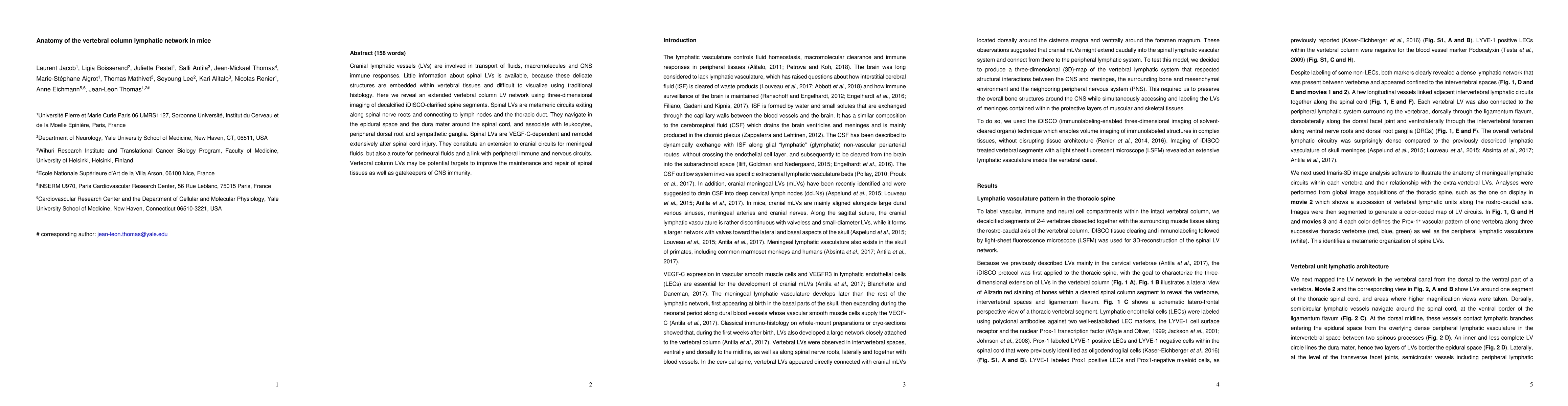
Cranial lymphatic vessels (LVs) are involved in transport of fluids, macromolecules and CNS immune responses. Little information about spinal LVs is available, because these delicate structures are embedded within vertebral tissues and difficult to visualize using traditional histology. Here we reveal an extended vertebral column LV network using three-dimensional imaging of decalcified iDISCO-clarified spine segments. Spinal LVs are metameric circuits exiting along spinal nerve roots and connecting to lymph nodes and the thoracic duct. They navigate in the epidural space and the dura mater around the spinal cord, and associate with leukocytes, peripheral dorsal root and sympathetic ganglia. Spinal LVs are VEGF-C-dependent and remodel extensively after spinal cord injury. They constitute an extension to cranial circuits for meningeal fluids, but also a route for perineural fluids and a link with peripheral immune and nervous circuits. Vertebral column LVs may be potential targets to improve the maintenance and repair of 32 spinal tissues as well as gatekeepers of CNS immunity.
AI Key Findings
Get AI-generated insights about this paper's methodology, results, and significance.
Paper Details
PDF Preview
Key Terms
Citation Network
Current paper (gray), citations (green), references (blue)
Display is limited for performance on very large graphs.
Similar Papers
Found 4 papersDevelopment of the Lymphatic System in the 4D XCAT Phantom
Arman Rahmim, Carlos F. Uribe, Roberto Fedrigo et al.
| Title | Authors | Year | Actions |
|---|
Comments (0)