Angle-resolved X-ray photoemission electron microscopy
Publication
Metrics
AI Quick Summary
This paper provides an overview of synchrotron-based angle-resolved X-ray photoemission electron microscopy (ARPES) and its applications in imaging the local electronic structure of materials. It demonstrates a darkfield XPEEM method for high-resolution mapping and showcases its effectiveness in imaging domains in a model oxide structure.
Paper Preview
Abstract
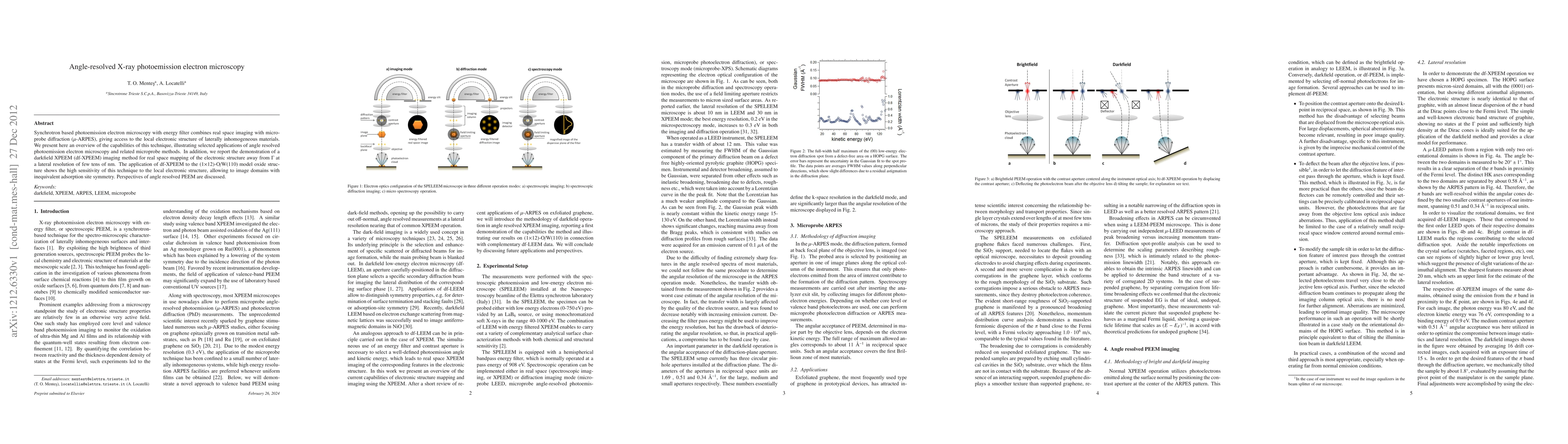
Synchrotron based photoemission electron microscopy with energy filter combines real space imaging with microprobe diffraction ($\mu$-ARPES), giving access to the local electronic structure of laterally inhomogeneous materials. We present here an overview of the capabilities of this technique, illustrating selected applications of angle resolved photoemission electron microscopy and related microprobe methods. In addition, we report the demonstration of a darkfield XPEEM (df-XPEEM) imaging method for real space mapping of the electronic structure away from $\Gamma$ at a lateral resolution of few tens of nm. The application of df-XPEEM to the (1$\times$12)-O/W(110) model oxide structure shows the high sensitivity of this technique to the local electronic structure, allowing to image domains with inequivalent adsorption site symmetry. Perspectives of angle resolved PEEM are discussed.
AI Key Findings
Get AI-generated insights about this paper's methodology, results, significance, and more — seven facets brought into focus.
Impact
Paper Details
PDF Preview
Key Terms
Citation Network
Current paper (gray), citations (green), references (blue)
Display is limited for performance on very large graphs.
Discussion 0