Anisotropic Contrast Optical Microscope
Publication
Metrics
AI Quick Summary
A new optical microscope reveals contrast in Mueller matrix images of thin specimens within anisotropic object planes, improving sensitivity for detecting organic mass by 4 orders of magnitude.
Paper Preview
Abstract
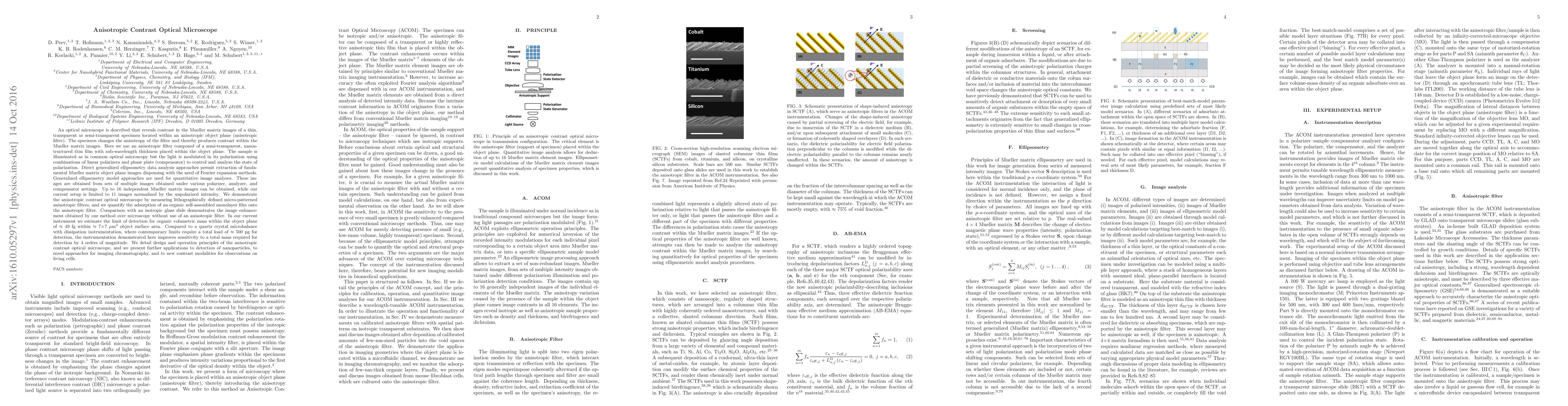
An optical microscope is described that reveals contrast in the Mueller matrix images of a thin, transparent or semi-transparent specimen located within an anisotropic object plane (anisotropic filter). The specimen changes the anisotropy of the filter and thereby produces contrast within the Mueller matrix images. Here we use an anisotropic filter composed of a semi-transparent, nanostructured thin film with sub-wavelength thickness placed within the object plane. The sample is illuminated as in common optical microscopy but the light is modulated in its polarization using combinations of linear polarizers and phase plate (compensator) to control and analyze the state of polarization. Direct generalized ellipsometry data analysis approaches permit extraction of fundamental Mueller matrix object plane images dispensing with the need of Fourier expansion methods. Generalized ellipsometry model approaches are used for quantitative image analyses. We demonstrate the anisotropic contrast optical microscope by measuring lithographically defined micro-patterned anisotropic filters, and we quantify the adsorption of an organic self-assembled monolayer film onto the anisotropic filter. In our current instrument we estimate the limit of detection for organic volumetric mass within the object plane of $\approx$ 49 fg within $\approx$ 7$\times$7~$\mu$m$^2$ object surface area. Compared to a quartz crystal microbalance with dissipation instrumentation, where contemporary limits require a total load of $\approx$ 500~pg for detection, the instrumentation demonstrated here improves sensitivity to a total mass required for detection by 4 orders of magnitude. We present further applications to detection of nanoparticles, to novel approaches for imaging chromatography, and to new contrast modalities for observations on living cells.
AI Key Findings
Get AI-generated insights about this paper's methodology, results, significance, and more — seven facets brought into focus.
Impact
Paper Details
PDF Preview
Key Terms
Citation Network
Current paper (gray), citations (green), references (blue)
Display is limited for performance on very large graphs.
Discussion 0