The research utilized a stable copolymer-in-oil base material to create anthropomorphic structures with optimized dyes mimicking blood optical absorption across a wide spectral range. 3D-printed phantom moulds from an MRI of a human forearm were used for shaping, and reflectance hyperspectral imaging (HSI) and photoacoustic tomography (PAT) were employed for image acquisition.
Anthropomorphic tissue-mimicking phantoms for oximetry validation in multispectral optical imaging
Publication
Metrics
AI Quick Summary
This paper introduces anthropomorphic tissue-mimicking phantoms designed to validate multispectral optical imaging methods for blood oxygenation (sO$_2$) assessment. The phantoms, created using stable copolymer-in-oil material and optimized dyes, correlate well with hyperspectral imaging and photoacoustic tomography data, offering a reliable tool for sO$_2$ biomarker validation.
Quick Answers
What is "Anthropomorphic tissue-mimicking phantoms for oximetry validation in multispectral optical imaging" about?
This paper introduces anthropomorphic tissue-mimicking phantoms designed to validate multispectral optical imaging methods for blood oxygenation (sO$_2$) assessment. The phantoms, created using stable copolymer-in-oil material and optimized dyes, correlate well with hyperspectral imaging and photoacoustic tomography data, offering a reliable tool for sO$_2$ biomarker validation.
What methodology did the authors use?
The research utilized a stable copolymer-in-oil base material to create anthropomorphic structures with optimized dyes mimicking blood optical absorption across a wide spectral range. 3D-printed phantom moulds from an MRI of a human forearm were used for shaping, and reflectance hyperspectral imaging (HSI) and photoacoustic tomography (PAT) were employed for image acquisition. More in Methodology →
What are the key results?
10 forearm phantoms with vessel-like structures and 5 distinct sO2 levels (0-100%) were fabricated. — Measured absorption spectra of the material correlated well with HSI and PAT data, with Pearson correlation coefficient consistently above 0.8. More in Key Results →
Why is this work significant?
These anthropomorphic tissue-mimicking phantoms offer a robust tool for developing, standardizing, and validating optical imaging of blood oxygenation (sO2) in clinical settings. More in Significance →
What are the main limitations?
The study did not explore the phantoms' performance under various environmental conditions (e.g., temperature, pressure). — The research focused on a single body part (forearm), and further validation on other anatomical regions is needed. More in Limitations →
Abstract
Significance: Optical imaging of blood oxygenation (sO$_2$) can be achieved based on the differential absorption spectra of oxy- and deoxy-haemoglobin. A key challenge in realising clinical validation of the sO$_2$ biomarkers is the absence of reliable sO$_2$ reference standards, including test objects. Aim: To enable quantitative testing of multispectral imaging methods for assessment of sO$_2$ by introducing anthropomorphic phantoms with appropriate tissue-mimicking optical properties. 
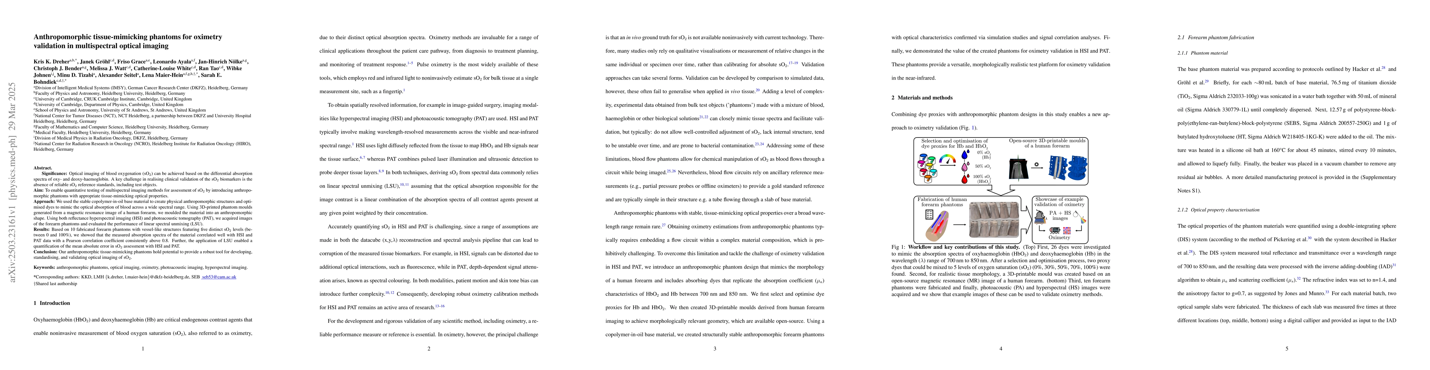
Approach: We used the stable copolymer-in-oil base material to create physical anthropomorphic structures and optimised dyes to mimic the optical absorption of blood across a wide spectral range. Using 3D-printed phantom moulds generated from a magnetic resonance image of a human forearm, we moulded the material into an anthropomorphic shape. Using both reflectance hyperspectral imaging (HSI) and photoacoustic tomography (PAT), we acquired images of the forearm phantoms and evaluated the performance of linear spectral unmixing (LSU). Results: Based on 10 fabricated forearm phantoms with vessel-like structures featuring five distinct sO$_2$ levels (between 0 and 100%), we showed that the measured absorption spectra of the material correlated well with HSI and PAT data with a Pearson correlation coefficient consistently above 0.8. Further, the application of LSU enabled a quantification of the mean absolute error in sO$_2$ assessment with HSI and PAT. Conclusion: Our anthropomorphic tissue-mimicking phantoms hold potential to provide a robust tool for developing, standardising, and validating optical imaging of sO$_2$.
Key Findings, in focus
Seven facets of this paper, analysed and brought into focus by AI.
These anthropomorphic tissue-mimicking phantoms offer a robust tool for developing, standardizing, and validating optical imaging of blood oxygenation (sO2) in clinical settings.
- 10 forearm phantoms with vessel-like structures and 5 distinct sO2 levels (0-100%) were fabricated.
- Measured absorption spectra of the material correlated well with HSI and PAT data, with Pearson correlation coefficient consistently above 0.8.
- Linear spectral unmixing (LSU) enabled quantification of mean absolute error in sO2 assessment with HSI and PAT.
These anthropomorphic tissue-mimicking phantoms offer a robust tool for developing, standardizing, and validating optical imaging of blood oxygenation (sO2) in clinical settings.
The introduction of anthropomorphic phantoms with tailored optical properties for sO2 assessment in multispectral optical imaging.
This work presents a novel approach to creating realistic test objects for validating sO2 biomarkers in optical imaging, addressing the absence of reliable reference standards.
- The study did not explore the phantoms' performance under various environmental conditions (e.g., temperature, pressure).
- The research focused on a single body part (forearm), and further validation on other anatomical regions is needed.
- Investigate phantom performance under diverse environmental conditions.
- Expand validation to other anatomical regions for broader applicability.
Impact
Authors
PDF Preview
Citation Network
Current paper (gray), citations (green), references (blue)
Display is limited for performance on very large graphs.
Discussion 0