APIS: A paired CT-MRI dataset for ischemic stroke segmentation challenge
Publication
Metrics
AI Quick Summary
APIS is a novel paired CT-MRI dataset for ischemic stroke segmentation, aiming to improve diagnosis by leveraging complementary data from non-contrast CT and diffusion-weighted MRI. Despite deep learning approaches, segmenting ischemic strokes from CT remains challenging, highlighting the dataset's potential to advance research.
Paper Preview
Abstract
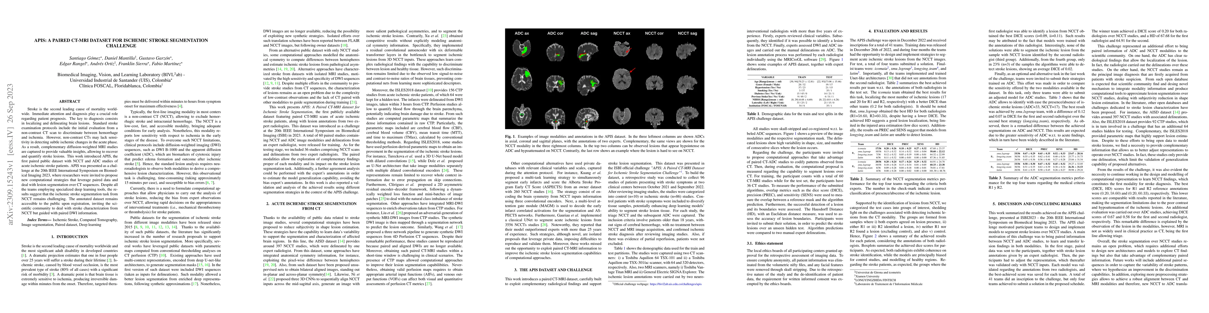
Stroke is the second leading cause of mortality worldwide. Immediate attention and diagnosis play a crucial role regarding patient prognosis. The key to diagnosis consists in localizing and delineating brain lesions. Standard stroke examination protocols include the initial evaluation from a non-contrast CT scan to discriminate between hemorrhage and ischemia. However, non-contrast CTs may lack sensitivity in detecting subtle ischemic changes in the acute phase. As a result, complementary diffusion-weighted MRI studies are captured to provide valuable insights, allowing to recover and quantify stroke lesions. This work introduced APIS, the first paired public dataset with NCCT and ADC studies of acute ischemic stroke patients. APIS was presented as a challenge at the 20th IEEE International Symposium on Biomedical Imaging 2023, where researchers were invited to propose new computational strategies that leverage paired data and deal with lesion segmentation over CT sequences. Despite all the teams employing specialized deep learning tools, the results suggest that the ischemic stroke segmentation task from NCCT remains challenging. The annotated dataset remains accessible to the public upon registration, inviting the scientific community to deal with stroke characterization from NCCT but guided with paired DWI information.
AI Key Findings
Get AI-generated insights about this paper's methodology, results, significance, and more — seven facets brought into focus.
Impact
Paper Details
Authors
PDF Preview
Key Terms
Citation Network
Current paper (gray), citations (green), references (blue)
Display is limited for performance on very large graphs.
Discussion 0