Artificial neural networks for 3D cell shape recognition from confocal images
Publication
Metrics
AI Quick Summary
This paper introduces a dual-stage neural network for 3D cell shape recognition from confocal microscopy images, tested on red blood cells from healthy donors and patients with congenital blood disease, aiming to provide unbiased diagnostic and theragnostic classification.
Paper Preview
Abstract
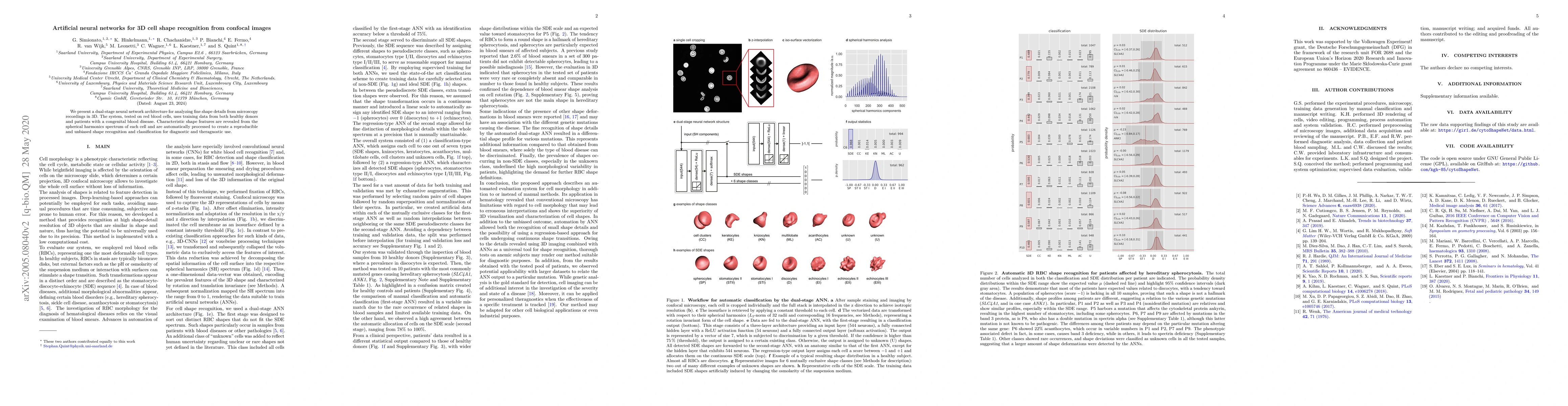
We present a dual-stage neural network architecture for analyzing fine shape details from microscopy recordings in 3D. The system, tested on red blood cells, uses training data from both healthy donors and patients with a congenital blood disease. Characteristic shape features are revealed from the spherical harmonics spectrum of each cell and are automatically processed to create a reproducible and unbiased shape recognition and classification for diagnostic and theragnostic use.
AI Key Findings
Get AI-generated insights about this paper's methodology, results, significance, and more — seven facets brought into focus.
Impact
Paper Details
PDF Preview
Key Terms
Citation Network
Current paper (gray), citations (green), references (blue)
Display is limited for performance on very large graphs.
Discussion 0