Publication
Metrics
AI Quick Summary
This study assesses bilateral neurovascular bundles' function in prostate radiotherapy patients using pulsed wave Doppler ultrasound, finding significant differences in Doppler waveform features that could guide NVB-sparing radiotherapy to reduce erectile dysfunction. The proposed method may enhance sexual function preservation and overall patient well-being.
Paper Preview
Abstract
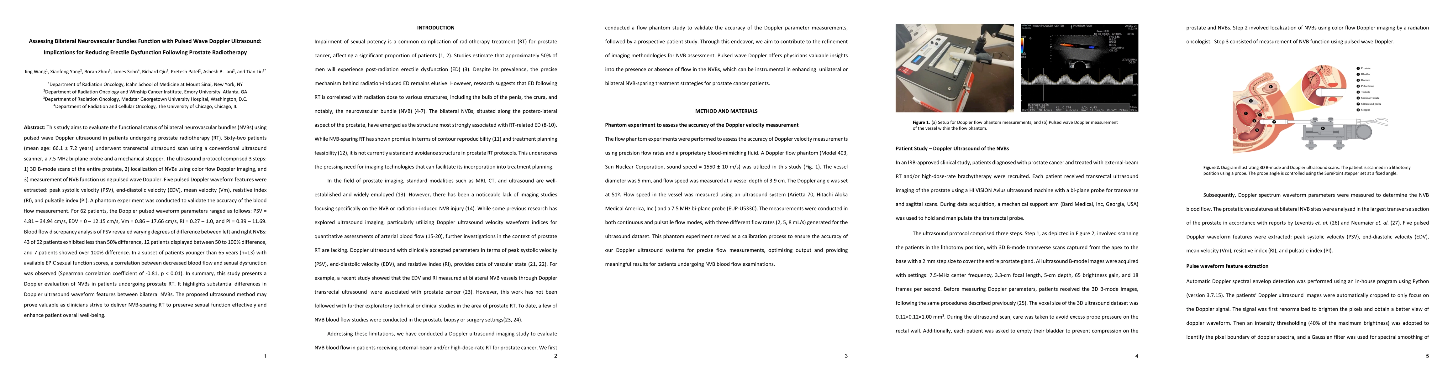
This study aims to evaluate the functional status of bilateral neurovascular bundles (NVBs) using pulsed wave Doppler ultrasound in patients undergoing prostate radiotherapy (RT). Sixty-two patients (mean age: 66.1 +/- 7.2 years) underwent transrectal ultrasound scan using a conventional ultrasound scanner, a 7.5 MHz bi-plane probe and a mechanical stepper. The ultrasound protocol comprised 3 steps: 1) 3D B-mode scans of the entire prostate, 2) localization of NVBs using color flow Doppler imaging, and 3) measurement of NVB function using pulsed wave Doppler. Five pulsed Doppler waveform features were extracted: peak systolic velocity (PSV), end-diastolic velocity (EDV), mean velocity (Vm), resistive index (RI), and pulsatile index (PI). In summary, this study presents a Doppler evaluation of NVBs in patients undergoing prostate RT. It highlights substantial differences in Doppler ultrasound waveform features between bilateral NVBs. The proposed ultrasound method may prove valuable as clinicians strive to deliver NVB-sparing RT to preserve sexual function effectively and enhance patients' overall well-being.
AI Key Findings
Get AI-generated insights about this paper's methodology, results, significance, and more — seven facets brought into focus.
Impact
Paper Details
Authors
PDF Preview
Key Terms
Citation Network
Current paper (gray), citations (green), references (blue)
Display is limited for performance on very large graphs.
Discussion 0