Assessing glaucoma in retinal fundus photographs using Deep Feature Consistent Variational Autoencoders
Publication
Metrics
AI Quick Summary
This paper proposes a Deep Feature Consistent Variational Autoencoder (DFC-VAE) to detect glaucoma in retinal fundus photographs. The model learns to reconstruct optic disc images and provides a small latent space that effectively discriminates between normal and glaucoma eyes, achieving an AUC of 0.885.
Paper Preview
Abstract
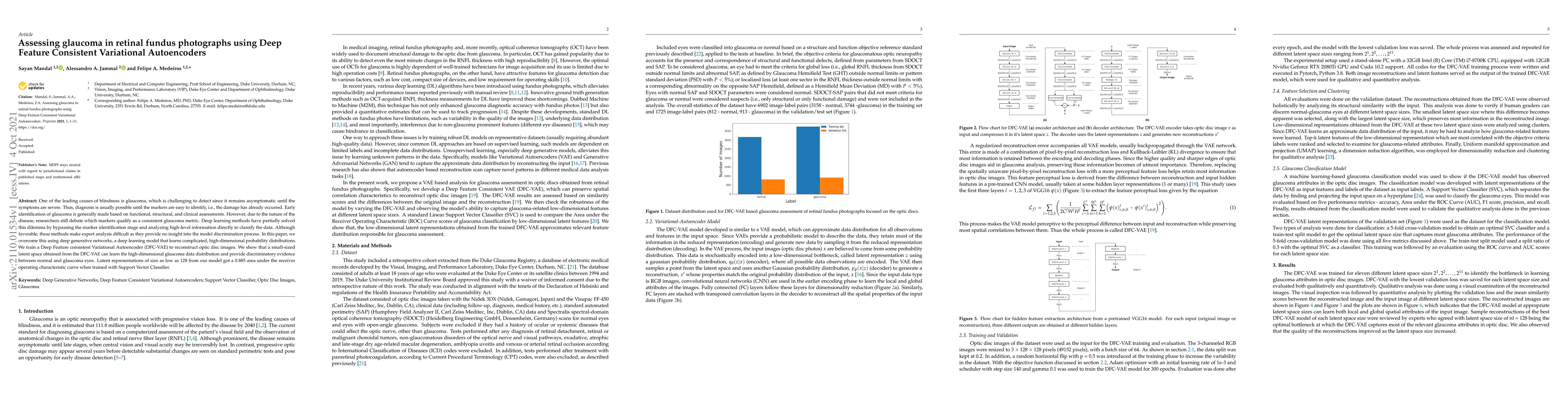
One of the leading causes of blindness is glaucoma, which is challenging to detect since it remains asymptomatic until the symptoms are severe. Thus, diagnosis is usually possible until the markers are easy to identify, i.e., the damage has already occurred. Early identification of glaucoma is generally made based on functional, structural, and clinical assessments. However, due to the nature of the disease, researchers still debate which markers qualify as a consistent glaucoma metric. Deep learning methods have partially solved this dilemma by bypassing the marker identification stage and analyzing high-level information directly to classify the data. Although favorable, these methods make expert analysis difficult as they provide no insight into the model discrimination process. In this paper, we overcome this using deep generative networks, a deep learning model that learns complicated, high-dimensional probability distributions. We train a Deep Feature consistent Variational Autoencoder (DFC-VAE) to reconstruct optic disc images. We show that a small-sized latent space obtained from the DFC-VAE can learn the high-dimensional glaucoma data distribution and provide discriminatory evidence between normal and glaucoma eyes. Latent representations of size as low as 128 from our model got a 0.885 area under the receiver operating characteristic curve when trained with Support Vector Classifier.
AI Key Findings
Get AI-generated insights about this paper's methodology, results, significance, and more — seven facets brought into focus.
Impact
Paper Details
Authors
PDF Preview
Key Terms
Citation Network
Current paper (gray), citations (green), references (blue)
Display is limited for performance on very large graphs.
Discussion 0