Asymmetric-detection time-stretch optical microscopy (ATOM) for ultrafast high-contrast cellular imaging in flow
Publication
Metrics
AI Quick Summary
Asymmetric-detection time-stretch optical microscopy (ATOM) enables ultrafast, high-contrast cellular imaging in flow, achieving label-free imaging at speeds up to 10 m/s and throughputs of ~100,000 cells/sec while maintaining morphological resolution and sensitivity. ATOM separates enhanced phase-gradient and absorption contrast, potentially revolutionizing cellular assay and imaging flow cytometry.
Paper Preview
Abstract
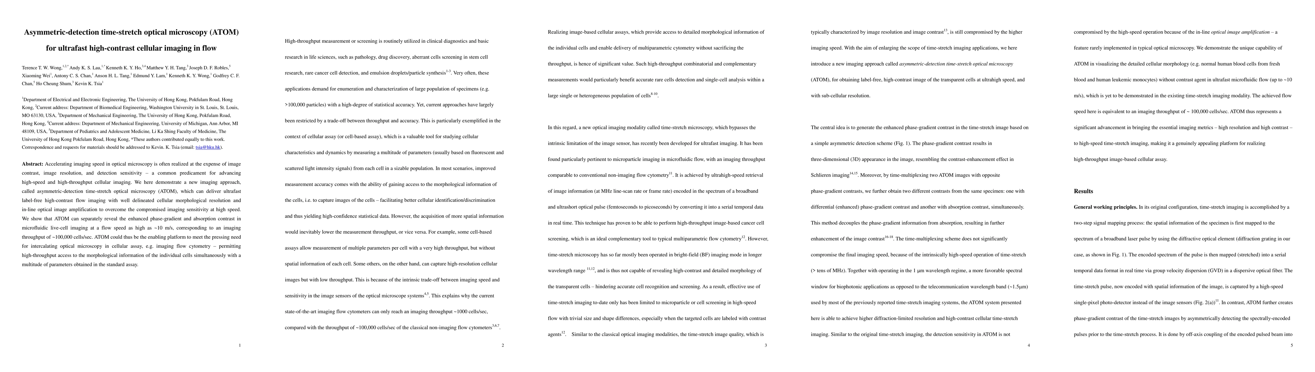
Accelerating imaging speed in optical microscopy is often realized at the expense of image contrast, image resolution, and detection sensitivity- a common predicament for advancing high-speed and high-throughput cellular imaging. We here demonstrate a new imaging approach, called asymmetric-detection time-stretch optical microscopy (ATOM), which can deliver ultrafast label-free high-contrast flow imaging with well delineated cellular morphological resolution and in-line optical image amplification to overcome the compromised imaging sensitivity at high speed. We show that ATOM can separately reveal the enhanced phase-gradient and absorption contrast in microfluidic live-cell imaging at a flow speed as high as ~10 m/s, corresponding to an imaging throughput of ~100,000 cells/sec. ATOM could thus be the enabling platform to meet the pressing need for intercalating optical microscopy in cellular assay, e.g. imaging flow cytometry- permitting high-throughput access to the morphological information of the individual cells simultaneously with a multitude of parameters obtained in the standard assay.
AI Key Findings
Get AI-generated insights about this paper's methodology, results, significance, and more — seven facets brought into focus.
Impact
Paper Details
PDF Preview
Key Terms
Citation Network
Current paper (gray), citations (green), references (blue)
Display is limited for performance on very large graphs.
Discussion 0