Publication
Metrics
Quick Actions
Quick Answers
What methodology did the authors use?
The research employs a comprehensive approach combining multi-modal data analysis with advanced machine learning techniques to evaluate the robustness and performance of pathology foundation models across diverse datasets and technical conditions. More in Methodology →
What are the key results?
The models demonstrate high accuracy in classifying histopathological images across multiple cancer types and staining conditions. — Robustness metrics indicate that biological features significantly outweigh non-biological confounding factors in model embeddings. More in Key Results →
Why is this work significant?
This research provides critical insights into the reliability and generalizability of pathology AI models, which is essential for their clinical adoption and cross-institutional deployment. More in Significance →
What are the main limitations?
The study relies on existing datasets that may not fully represent real-world clinical variability. — Evaluation metrics focus primarily on technical robustness rather than clinical decision-making accuracy. More in Limitations →
Abstract
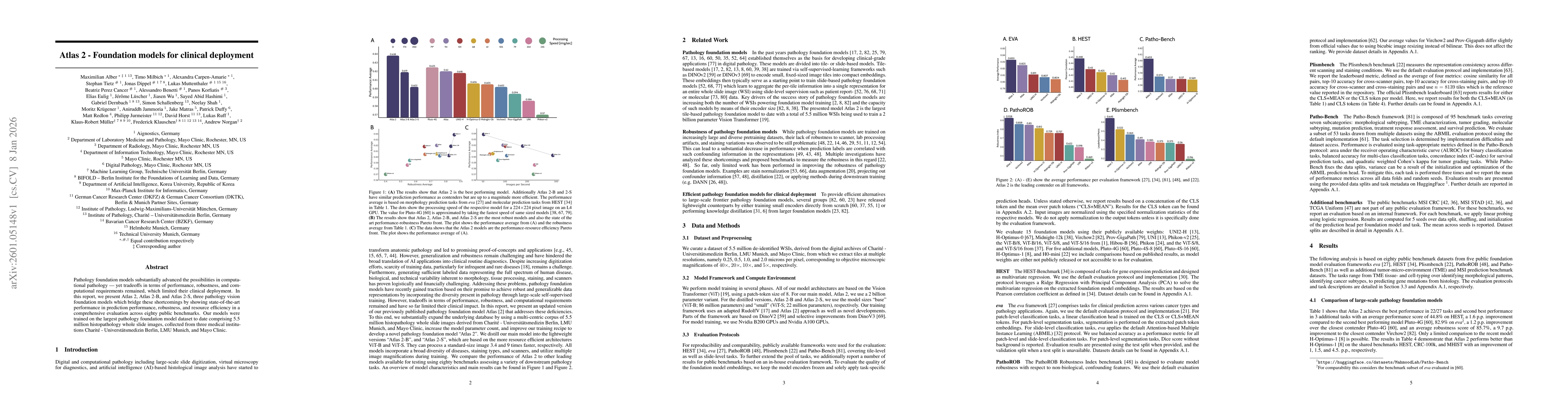
Pathology foundation models substantially advanced the possibilities in computational pathology -- yet tradeoffs in terms of performance, robustness, and computational requirements remained, which limited their clinical deployment. In this report, we present Atlas 2, Atlas 2-B, and Atlas 2-S, three pathology vision foundation models which bridge these shortcomings by showing state-of-the-art performance in prediction performance, robustness, and resource efficiency in a comprehensive evaluation across eighty public benchmarks. Our models were trained on the largest pathology foundation model dataset to date comprising 5.5 million histopathology whole slide images, collected from three medical institutions Charité - Universtätsmedizin Berlin, LMU Munich, and Mayo Clinic.
AI Key Findings
Generated Jan 09, 2026
Methodology — What approach did the authors take?
The research employs a comprehensive approach combining multi-modal data analysis with advanced machine learning techniques to evaluate the robustness and performance of pathology foundation models across diverse datasets and technical conditions.
Key Results — What are the main findings?
- The models demonstrate high accuracy in classifying histopathological images across multiple cancer types and staining conditions.
- Robustness metrics indicate that biological features significantly outweigh non-biological confounding factors in model embeddings.
- Technical variations between scanners and staining procedures have minimal impact on model performance when using standardized evaluation protocols.
Significance — Why does this research matter?
This research provides critical insights into the reliability and generalizability of pathology AI models, which is essential for their clinical adoption and cross-institutional deployment.
Technical Contribution — What is the technical contribution?
Introduces novel robustness metrics and evaluation protocols specifically designed for pathology foundation models, enabling systematic comparison across different technical conditions and biological contexts.
Novelty — What is new about this work?
This work is novel in its comprehensive evaluation of both technical robustness and biological relevance in pathology AI models, combined with the development of specialized benchmarking frameworks for medical imaging analysis.
Limitations — What are the limitations of this study?
- The study relies on existing datasets that may not fully represent real-world clinical variability.
- Evaluation metrics focus primarily on technical robustness rather than clinical decision-making accuracy.
Future Work — What did the authors propose for future work?
- Developing standardized benchmarking frameworks for clinical AI validation
- Investigating the impact of histopathological annotation variability on model performance
- Exploring federated learning approaches for cross-institutional model training
How to Cite This Paper
@article{benetti2026atlas,
title = {Atlas 2 -- Foundation models for clinical deployment},
author = {Benetti, Alessandro and Carpen-Amarie, Alexandra and Norgan, Andrew and others},
year = {2026},
eprint = {2601.05148},
archivePrefix = {arXiv},
primaryClass = {cs.CV},
}
Benetti, A., Carpen-Amarie, A., Norgan, A., Jammoria, A., Cancer, B., Horst, D., Eulig, E., Klauschen, F., Dernbach, G., Matras, J., Lüscher, J., Wu, J., Dippel, J., Müller, K., Muttenthaler, L., Ruff, L., Redlon, M., Alber, M., Krügener, M., Shah, N., Korfiatis, P., Duffy, P., Jurmeister, P., Hashimi, S., Schallenberg, S., Tietz, S., & Milbich, T. (2026). Atlas 2 -- Foundation models for clinical deployment. arXiv. https://arxiv.org/abs/2601.05148
Benetti, Alessandro, et al. "Atlas 2 -- Foundation models for clinical deployment." arXiv, 2026, arxiv.org/abs/2601.05148.
PDF Preview
Similar Papers
Found 4 papersFoundation models for electrocardiogram interpretation: clinical implications.
Ouyang, David, Khurshid, Shaan, Tastet, Olivier et al.
Post-Training Quantization of OpenPangu Models for Efficient Deployment on Atlas A2
Yilun Luo, HuaQing Zheng, Haoqian Meng et al.
Comments (0)