Atomic force microscopy (AFM) study of thick lamellar stacks of phospholipid bilayers
Publication
Metrics
AI Quick Summary
This study uses Atomic Force Microscopy (AFM) to investigate thick stacks of DMPC phospholipid bilayers, determining their compressional modulus and rupture force across different phases. AFM reveals pronounced ripples in the gel phase, with ripple period increasing near the main phase transition and under varying osmotic pressure.
Paper Preview
Abstract
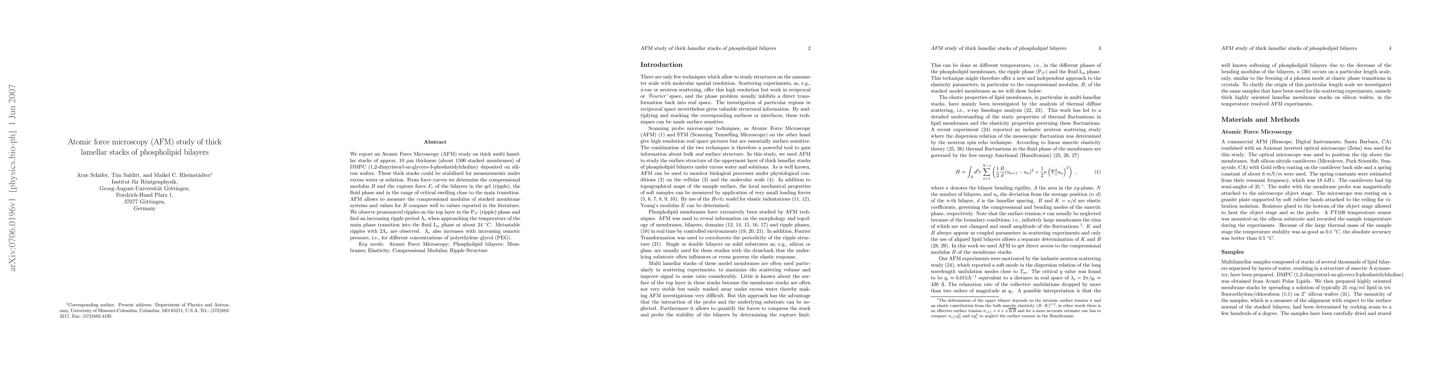
We report an Atomic Force Microscopy (AFM) study on thick multi lamellar stacks of approx. 10 mum thickness (about 1500 stacked membranes) of DMPC (1,2-dimyristoyl-sn-glycero-3-phoshatidylcholine) deposited on silicon wafers. These thick stacks could be stabilized for measurements under excess water or solution. From force curves we determine the compressional modulus B and the rupture force F_r of the bilayers in the gel (ripple), the fluid phase and in the range of critical swelling close to the main transition. AFM allows to measure the compressional modulus of stacked membrane systems and values for B compare well to values reported in the literature. We observe pronounced ripples on the top layer in the Pbeta' (ripple) phase and find an increasing ripple period Lambda_r when approaching the temperature of the main phase transition into the fluid Lalpha phase at about 24 C. Metastable ripples with 2Lambda_r are observed. Lambda_r also increases with increasing osmotic pressure, i.e., for different concentrations of polyethylene glycol (PEG).
AI Key Findings
Get AI-generated insights about this paper's methodology, results, significance, and more — seven facets brought into focus.
Impact
Paper Details
PDF Preview
Key Terms
Citation Network
Current paper (gray), citations (green), references (blue)
Display is limited for performance on very large graphs.
Discussion 0