Summary
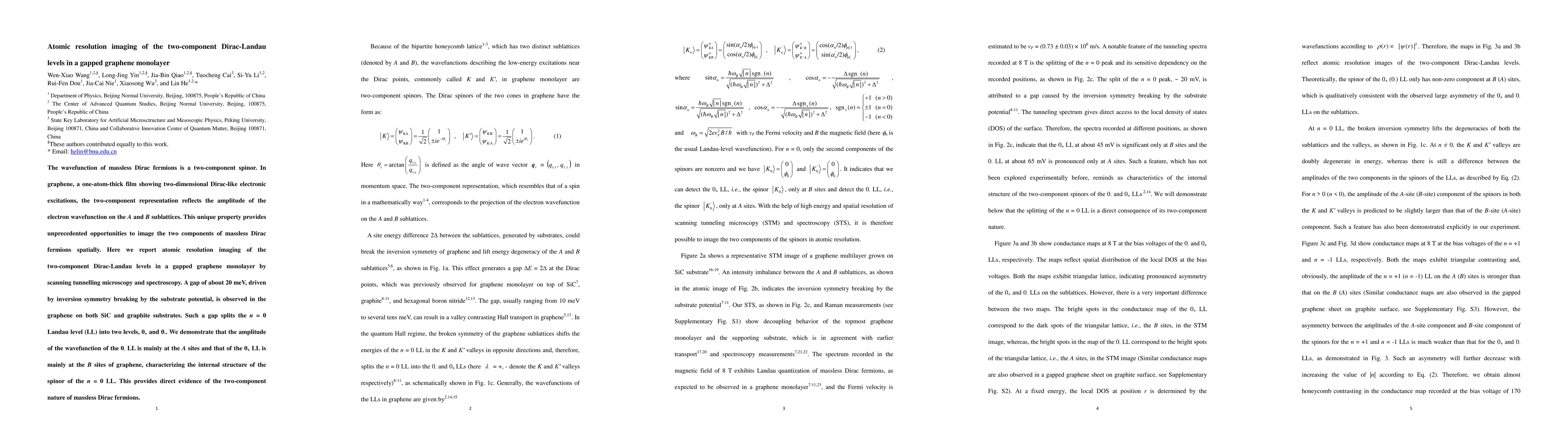
The wavefunction of massless Dirac fermions is a two-component spinor. In graphene, a one-atom-thick film showing two-dimensional Dirac-like electronic excitations, the two-component representation reflects the amplitude of the electron wavefunction on the A and B sublattices. This unique property provides unprecedented opportunities to image the two components of massless Dirac fermions spatially. Here we report atomic resolution imaging of the two-component Dirac-Landau levels in a gapped graphene monolayer by scanning tunnelling microscopy and spectroscopy. A gap of about 20 meV, driven by inversion symmetry breaking by the substrate potential, is observed in the graphene on both SiC and graphite substrates. Such a gap splits the n = 0 Landau level (LL) into two levels, 0+ and 0-. We demonstrate that the amplitude of the wavefunction of the 0- LL is mainly at the A sites and that of the 0+ LL is mainly at the B sites of graphene, characterizing the internal structure of the spinor of the n = 0 LL. This provides direct evidence of the two-component nature of massless Dirac fermions.
AI Key Findings
Get AI-generated insights about this paper's methodology, results, and significance.
Paper Details
PDF Preview
Key Terms
Citation Network
Current paper (gray), citations (green), references (blue)
Display is limited for performance on very large graphs.
| Title | Authors | Year | Actions |
|---|
Comments (0)