Atomic-Resolution Visualization and Doping Effects of Complex Structures in Intercalated Bilayer Graphene
Publication
Metrics
AI Quick Summary
Researchers used advanced microscopy and calculations to visualize atomic structures in bilayer graphene intercalated with FeCl3, discovering two distinct structures that affect the material's properties.
Paper Preview
Abstract
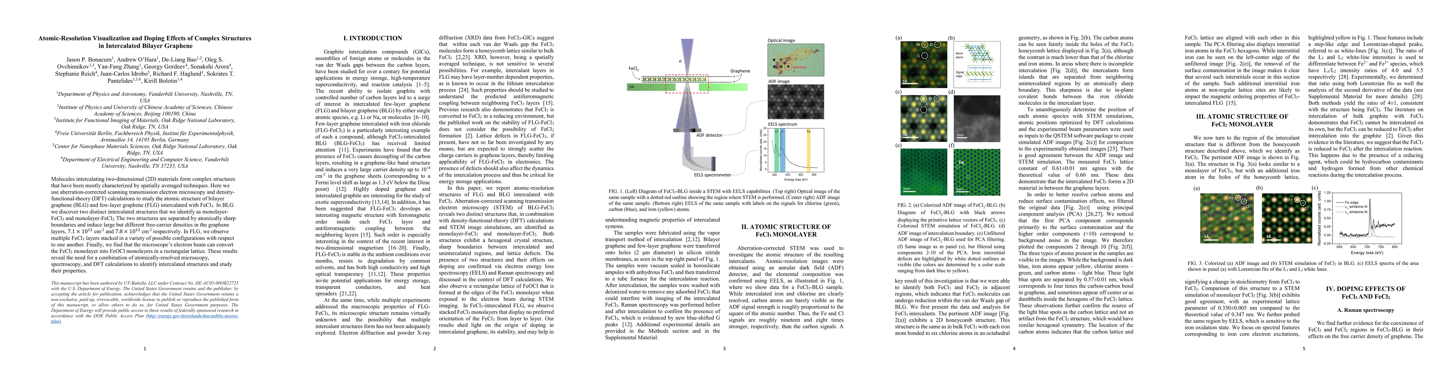
Molecules intercalating two-dimensional (2D) materials form complex structures that have been mostly characterized by spatially averaged techniques. Here we use aberration-corrected scanning transmission electron microscopy and density-functional-theory (DFT) calculations to study the atomic structure of bilayer graphene (BLG) and few-layer graphene (FLG) intercalated with FeCl$_3$. In BLG we discover two distinct intercalated structures that we identify as monolayer-FeCl$_3$ and monolayer-FeCl$_2$. The two structures are separated by atomically sharp boundaries and induce large but different free-carrier densities in the graphene layers, $7.1\times10^{13}$ cm$^{-2}$ and $7.1\times10^{13}$ cm$^{-2}$ respectively. In FLG, we observe multiple FeCl$_3$ layers stacked in a variety of possible configurations with respect to one another. Finally, we find that the microscope's electron beam can convert the FeCl$_3$ monolayer into FeOCl monolayers in a rectangular lattice. These results reveal the need for a combination of atomically-resolved microscopy, spectroscopy, and DFT calculations to identify intercalated structures and study their properties.
AI Key Findings
Get AI-generated insights about this paper's methodology, results, significance, and more — seven facets brought into focus.
Impact
Paper Details
PDF Preview
Key Terms
Citation Network
Current paper (gray), citations (green), references (blue)
Display is limited for performance on very large graphs.
Discussion 0