Authors
Summary
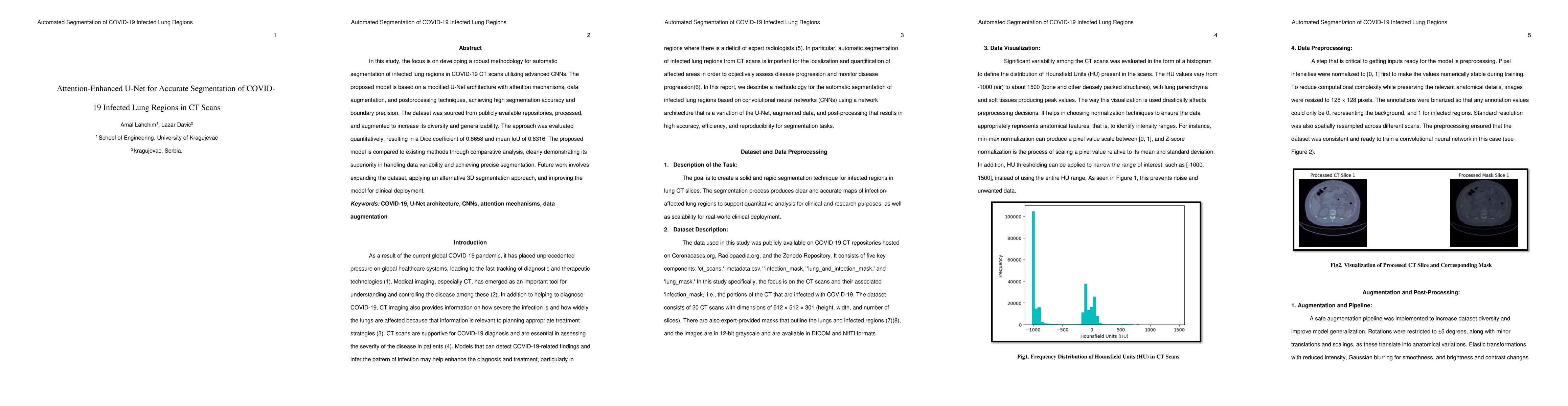
In this study, we propose a robust methodology for automatic segmentation of infected lung regions in COVID-19 CT scans using convolutional neural networks. The approach is based on a modified U-Net architecture enhanced with attention mechanisms, data augmentation, and postprocessing techniques. It achieved a Dice coefficient of 0.8658 and mean IoU of 0.8316, outperforming other methods. The dataset was sourced from public repositories and augmented for diversity. Results demonstrate superior segmentation performance. Future work includes expanding the dataset, exploring 3D segmentation, and preparing the model for clinical deployment.
AI Key Findings
Generated Jun 08, 2025
Methodology
The research proposes a method for automatic segmentation of infected lung regions in COVID-19 CT scans using a modified U-Net architecture enhanced with attention mechanisms, data augmentation, and postprocessing techniques.
Key Results
- Achieved a Dice coefficient of 0.8658 and mean IoU of 0.8316, outperforming other methods.
- Demonstrated superior segmentation performance compared to existing techniques.
Significance
This research is important as it provides a robust method for accurate segmentation of COVID-19 infected lung regions in CT scans, which can aid in diagnosis and treatment planning.
Technical Contribution
The main technical contribution is the attention-enhanced U-Net architecture for improved segmentation accuracy.
Novelty
This work is novel due to the integration of attention mechanisms into the U-Net architecture for enhanced performance in segmenting COVID-19 infected lung regions in CT scans.
Limitations
- The dataset was sourced from public repositories, which might limit diversity and representativeness.
- The study did not explore 3D segmentation despite its potential benefits.
Future Work
- Expanding the dataset for broader applicability and robustness.
- Exploring 3D segmentation for more comprehensive lung region analysis.
- Preparing the model for clinical deployment.
Paper Details
PDF Preview
Similar Papers
Found 4 papersCHS-Net: A Deep learning approach for hierarchical segmentation of COVID-19 infected CT images
Sonali Agarwal, Narinder Singh Punn
Automated Chest CT Image Segmentation of COVID-19 Lung Infection based on 3D U-Net
Dominik Müller, Frank Kramer, Iñaki Soto Rey
No citations found for this paper.
Comments (0)