This paper proposes an algorithmic method using a deep convolutional network to create a mitotic cell map, which aids in selecting the region with the highest mitotic activity for expert mitotic count determination, reducing variability in tumor grading. The method shows a strong correlation (r=0.936) with actual mitotic counts in a test set of 10 whole slide images.
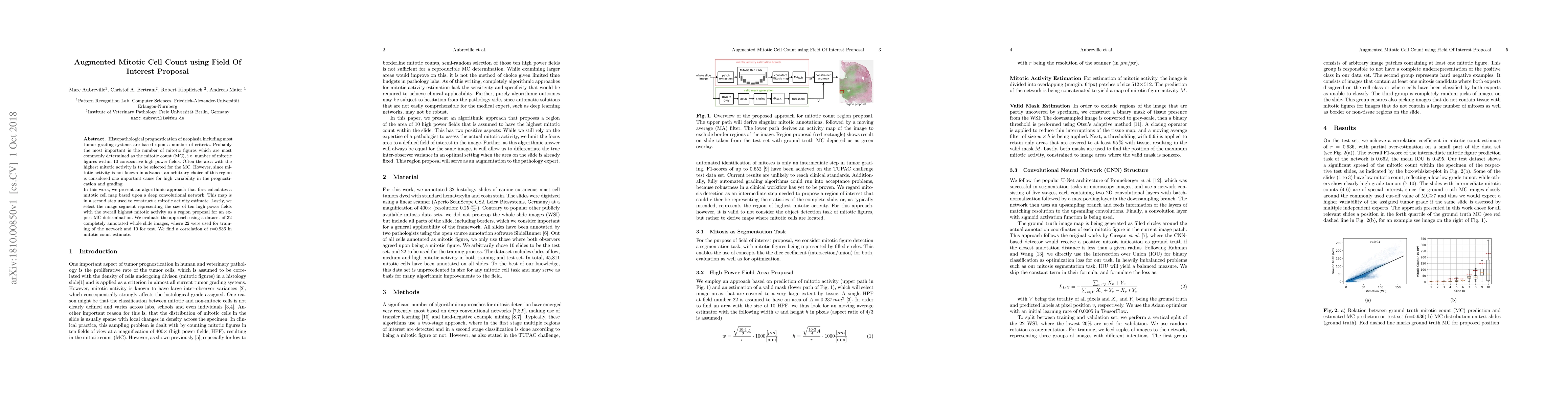
Histopathological prognostication of neoplasia including most tumor grading systems are based upon a number of criteria. Probably the most important is the number of mitotic figures which are most commonly determined as the mitotic count (MC), i.e. number of mitotic figures within 10 consecutive high power fields. Often the area with the highest mitotic activity is to be selected for the MC. However, since mitotic activity is not known in advance, an arbitrary choice of this region is considered one important cause for high variability in the prognostication and grading. In this work, we present an algorithmic approach that first calculates a mitotic cell map based upon a deep convolutional network. This map is in a second step used to construct a mitotic activity estimate. Lastly, we select the image segment representing the size of ten high power fields with the overall highest mitotic activity as a region proposal for an expert MC determination. We evaluate the approach using a dataset of 32 completely annotated whole slide images, where 22 were used for training of the network and 10 for test. We find a correlation of r=0.936 in mitotic count estimate.
Get AI-generated insights about this paper's methodology, results, and significance.
Current paper (gray), citations (green), references (blue)
Mert R. Sabuncu, Cagla Deniz Bahadir, Benjamin Liechty et al.
Jahanifar, M., Dawood, M., Zamanitajeddin, N. et al.
Title | Authors | Year | Actions |
---|---|---|---|
MiNuGAN: Dual Segmentation of Mitoses and Nuclei Using Conditional GANs on Multi-center Breast H&E Images
|
Salar Razavi, Fariba D. Khameneh, Hana Nouri, D. Androutsos, S. Done, A. Khademi
|
2022
|
View Paper |
A large-scale dataset for mitotic figure assessment on whole slide images of canine cutaneous mast cell tumor
|
C. Bertram, M. Aubreville, C. Marzahl, A. Maier, R. Klopfleisch
|
2019
|
View Paper |
Deep learning algorithms out-perform veterinary pathologists in detecting the mitotically most active tumor region
|
M. Aubreville, C. Bertram, C. Marzahl, C. Gurtner, M. Dettwiler, A. Schmidt, F. Bartenschlager, S. Merz, Marco Fragoso, O. Kershaw, R. Klopfleisch, A. Maier
|
2019
|
View Paper |
Field of Interest Prediction for Computer-Aided Mitotic Count
|
M. Aubreville, C. Bertram, C. Marzahl, C. Gurtner, M. Dettwiler, A. Schmidt, F. Bartenschlager, S. Merz, Marco Fragoso, O. Kershaw, R. Klopfleisch, A. Maier
|
2019
|
View Paper |
Field Of Interest Proposal for Augmented Mitotic Cell Count: Comparison of two Convolutional Networks
|
M. Aubreville, C. Bertram, R. Klopfleisch, A. Maier
|
2018
|
View Paper |
Comments (0)