Auto-context Convolutional Neural Network (Auto-Net) for Brain Extraction in Magnetic Resonance Imaging
Publication
Metrics
AI Quick Summary
This paper introduces an auto-context convolutional neural network (Auto-Net) for brain extraction from MRI scans, achieving superior accuracy on benchmark datasets and demonstrating effectiveness in extracting arbitrarily-oriented fetal brains without requiring 3D convolutions. The method uses posterior probability maps iteratively to refine brain segmentation.
Paper Preview
Abstract
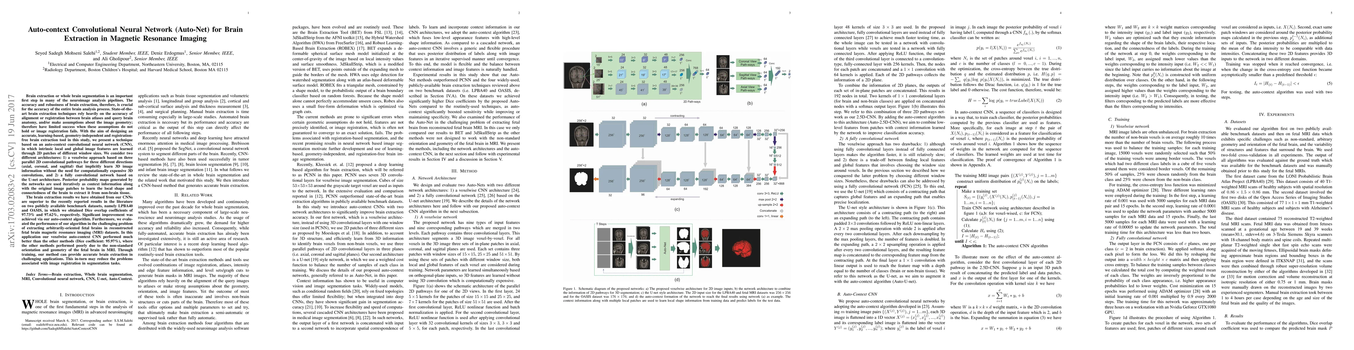
Brain extraction or whole brain segmentation is an important first step in many of the neuroimage analysis pipelines. The accuracy and robustness of brain extraction, therefore, is crucial for the accuracy of the entire brain analysis process. With the aim of designing a learning-based, geometry-independent and registration-free brain extraction tool in this study, we present a technique based on an auto-context convolutional neural network (CNN), in which intrinsic local and global image features are learned through 2D patches of different window sizes. In this architecture three parallel 2D convolutional pathways for three different directions (axial, coronal, and sagittal) implicitly learn 3D image information without the need for computationally expensive 3D convolutions. Posterior probability maps generated by the network are used iteratively as context information along with the original image patches to learn the local shape and connectedness of the brain, to extract it from non-brain tissue. The brain extraction results we have obtained from our algorithm are superior to the recently reported results in the literature on two publicly available benchmark datasets, namely LPBA40 and OASIS, in which we obtained Dice overlap coefficients of 97.42% and 95.40%, respectively. Furthermore, we evaluated the performance of our algorithm in the challenging problem of extracting arbitrarily-oriented fetal brains in reconstructed fetal brain magnetic resonance imaging (MRI) datasets. In this application our algorithm performed much better than the other methods (Dice coefficient: 95.98%), where the other methods performed poorly due to the non-standard orientation and geometry of the fetal brain in MRI. Our CNN-based method can provide accurate, geometry-independent brain extraction in challenging applications.
AI Key Findings
Get AI-generated insights about this paper's methodology, results, significance, and more — seven facets brought into focus.
Impact
Paper Details
PDF Preview
Key Terms
Citation Network
Current paper (gray), citations (green), references (blue)
Display is limited for performance on very large graphs.
Discussion 0