This study developed an automated clustering method using neural networks to discover human-interpretable dermatoscopic patterns from a large skin lesion dataset. The method resulted in fewer non-informative clusters and a higher proportion of clusters that matched known diagnostic patterns when using a compactness metric.
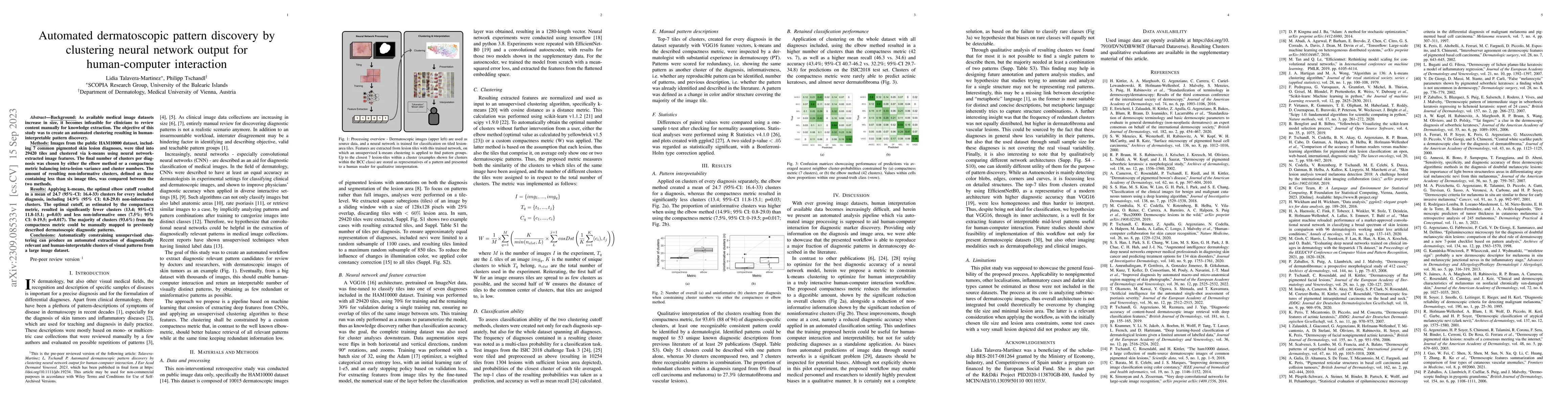
Background: As available medical image datasets increase in size, it becomes infeasible for clinicians to review content manually for knowledge extraction. The objective of this study was to create an automated clustering resulting in human-interpretable pattern discovery. Methods: Images from the public HAM10000 dataset, including 7 common pigmented skin lesion diagnoses, were tiled into 29420 tiles and clustered via k-means using neural network-extracted image features. The final number of clusters per diagnosis was chosen by either the elbow method or a compactness metric balancing intra-lesion variance and cluster numbers. The amount of resulting non-informative clusters, defined as those containing less than six image tiles, was compared between the two methods. Results: Applying k-means, the optimal elbow cutoff resulted in a mean of 24.7 (95%-CI: 16.4-33) clusters for every included diagnosis, including 14.9% (95% CI: 0.8-29.0) non-informative clusters. The optimal cutoff, as estimated by the compactness metric, resulted in significantly fewer clusters (13.4; 95%-CI 11.8-15.1; p=0.03) and less non-informative ones (7.5%; 95% CI: 0-19.5; p=0.017). The majority of clusters (93.6%) from the compactness metric could be manually mapped to previously described dermatoscopic diagnostic patterns. Conclusions: Automatically constraining unsupervised clustering can produce an automated extraction of diagnostically relevant and human-interpretable clusters of visual patterns from a large image dataset.
Get AI-generated insights about this paper's methodology, results, and significance.
Current paper (gray), citations (green), references (blue)
No citations found for this paper.
Title | Authors | Year | Actions |
---|---|---|---|
Improved diagnosis by automated macro‐ and micro‐anatomical region mapping of skin photographs
|
L. Amruthalingam, Philippe Gottfrois, A. Gonzalez Jimenez, B. Gökduman, M. Kunz, T. Koller, Dermanatomy Consortium, M. Pouly, A. Navarini
|
2022
|
View Paper |
Deep learning‐based classification of dermatological lesions given a limited amount of labelled data
|
S. Krammer, Y. Li, N. Jakob, A. S. Boehm, H. Wolff, P. Tang, Tobias Lasser, L. French, D. Hartmann
|
2022
|
View Paper |
Artificial intelligence for the automated single‐shot assessment of psoriasis severity
|
Takashi Okamoto, Masataka Kawai, Y. Ogawa, S. Shimada, T. Kawamura
|
2022
|
View Paper |
Dermoscopic Features Summarization and Comparison of Four Types of Cutaneous Vascular Anomalies
|
Jing Gao, Wenmin Fei, Changbing Shen, Xue Shen, Minghui Sun, Ning Xu, Qing Li, Cong Huang, Tingfang Zhang, Randy F. Ko, Yong Cui, Chunjun Yang
|
2021
|
View Paper |
Dermoscopic Predictors of Tumor Thickness in Cutaneous Melanoma: A Retrospective Analysis of 245 Melanomas
|
E. Rodríguez-Lomba, B. Lozano-Masdemont, L. M. Nieto-Benito, E. Hernández de la Torre, R. Suárez-Fernández, J. Avilés-Izquierdo
|
2021
|
View Paper |
Evaluating Deep Neural Networks Trained on Clinical Images in Dermatology with the Fitzpatrick 17k Dataset
|
Matthew Groh, Caleb Harris, L. Soenksen, Felix Lau, Rachel Han, Aerin Kim, A. Koochek, Omar Badri
|
2021
|
View Paper |
Dermatoscopic features of pigmented intraepidermal carcinoma on the head and neck
|
Mike Inskip, A. Cameron, B. N. Akay, M. Gorji, Simon P Clark, Nikita Rosendahl, Martelle Coetzer-Botha, H. Kittler, C. Rosendahl
|
2020
|
View Paper |
Human–computer collaboration for skin cancer recognition
|
P. Tschandl, C. Rinner, Z. Apalla, G. Argenziano, N. Codella, A. Halpern, M. Janda, A. Lallas, C. Longo, J. Malvehy, J. Malvehy, J. Paoli, J. Paoli, S. Puig, S. Puig, C. Rosendahl, H. Soyer, I. Zalaudek, H. Kittler
|
2020
|
View Paper |
Augment Intelligence Dermatology : Deep Neural Networks Empower Medical Professionals in Diagnosing Skin Cancer and Predicting Treatment Options for 134 Skin Disorders.
|
S. Han, I. Park, Sung Eun Chang, Woohyung Lim, M. Kim, Gyeong-Hun Park, J. Chae, C. Huh, J. Na
|
2020
|
View Paper |
Man against machine reloaded: performance of a market-approved convolutional neural network in classifying a broad spectrum of skin lesions in comparison with 96 dermatologists working under less artificial conditions.
|
H. Haenssle, C. Fink, F. Toberer, J. Winkler, W. Stolz, T. Deinlein, R. Hofmann-Wellenhof, A. Lallas, S. Emmert, T. Buhl, M. Zutt, A. Blum, M. S. Abassi, Luc Thomas, I. Tromme, P. Tschandl, A. Enk, A. Rosenberger, C. Alt, M. Bachelerie, Sonali Bajaj, Alise Balcere, S. Baricault, Clément Barthaux, Yvonne Beckenbauer, I. Bertlich, A. Blum, M. Bouthenet, S. Brassat, Philipp Marcel Buck, K. Buder-Bakhaya, Maria-Letizia Cappelletti, Cécile Chabbert, Julie De Labarthe, Eveline Decoster, T. Deinlein, Michèle Dobler, Daphnée Dumon, S. Emmert, Julie Gachon-Buffet, M. Gusarov, Franziska Hartmann, J. Hartmann, Anke Herrmann, I. Hoorens, E. Hulstaert, R. Karls, Andreea Kolonte, C. Kromer, A. Lallas, Céline Le Blanc Vasseux, A. Levy-Roy, P. Majenka, M. Marc, Veronique Martin Bourret, Nadège Michelet-Brunacci, C. Mitteldorf, Jean-Baptiste Paroissien, Camille Picard, Diana Plise, Valérie Reymann, Fabrice Ribeaudeau, P. Richez, Hélène Roche Plaine, D. Salik, E. Sattler, Sarah Schäfer, R. Schneiderbauer, Thierry Secchi, K. Talour, Lukas Trennheuser, Alexander Wald, Priscila Wölbing, P. Zukervar
|
2020
|
View Paper |
BCN20000: Dermoscopic Lesions in the Wild
|
M. Combalia, N. Codella, V. Rotemberg, Brian Helba, Verónica Vilaplana, O. Reiter, A. Halpern, S. Puig, J. Malvehy
|
2019
|
View Paper |
Standardization of dermoscopic terminology and basic dermoscopic parameters to evaluate in general dermatology (non‐neoplastic dermatoses): an expert consensus on behalf of the International Dermoscopy Society
|
E. Errichetti, I. Zalaudek, H. Kittler, Z. Apalla, G. Argenziano, R. Bakos, A. Blum, R. Braun, D. Ioannides, F. Lacarrubba, E. Lazaridou, C. Longo, G. Micali, E. Moscarella, J. Paoli, C. Papageorgiou, T. Russo, A. Scope, G. Stinco, Luc Thomas, R. Toncic, P. Tschandl, H. Cabo, A. Hallpern, R. Hofmann-Wellenhof, J. Malvehy, A. Marghoob, S. Menzies, G. Pellacani, S. Puig, H. Rabinovitz, L. Rudnicka, E. Vakirlis, P. Soyer, Wilhelm Stolz, Masaru Tanaka, A. Lallas
|
2019
|
View Paper |
SciPy 1.0: fundamental algorithms for scientific computing in Python
|
Pauli Virtanen, R. Gommers, T. Oliphant, Matt Haberland, Tyler Reddy, D. Cournapeau, Evgeni Burovski, Pearu Peterson, Warren Weckesser, Jonathan Bright, S. J. van der Walt, M. Brett, Joshua Wilson, K. Millman, N. Mayorov, Andrew R. J. Nelson, E. Jones, Robert Kern, Eric Larson, C. Carey, İlhan Polat, Yu Feng, Eric W. Moore, J. Vanderplas, D. Laxalde, Josef Perktold, R. Cimrman, Ian Henriksen, E. Quintero, Charles R. Harris, A. Archibald, Antônio H. Ribeiro, Fabian Pedregosa, P. van Mulbregt, Aditya Alessandro Pietro Alex Andreas Andreas Anthony Ant Vijaykumar Bardelli Rothberg Hilboll Kloeckner Sco, A. Vijaykumar, Alessandro Pietro Bardelli, Alex Rothberg, A. Hilboll, Andre Kloeckner, A. Scopatz, Antony Lee, Ariel S. Rokem, C. N. Woods, Chad Fulton, Charles Masson, C. Häggström, Clark Fitzgerald, D. Nicholson, David R. Hagen, D. Pasechnik, E. Olivetti, Eric Martin, Eric Wieser, Fabrice Silva, F. Lenders, Florian Wilhelm, G. Young, Gavin A. Price, G. Ingold, Gregory E. Allen, Gregory R. Lee, H. Audren, I. Probst, J. Dietrich, J. Silterra, James T. Webber, J. Slavič, J. Nothman, J. Buchner, Johannes Kulick, Johannes L. Schönberger, J. V. De Miranda Cardoso, J. Reimer, J. Harrington, Juan Rodríguez, Juan Nunez-Iglesias, Justin Kuczynski, K. Tritz, M. Thoma, M. Newville, Matthias Kümmerer, Maximilian Bolingbroke, Michael Tartre, M. Pak, Nathaniel J. Smith, N. Nowaczyk, Nikolay Shebanov, O. Pavlyk, P. A. Brodtkorb, Perry Lee, R. McGibbon, Roman Feldbauer, Sam Lewis, S. Tygier, Scott Sievert, S. Vigna, Stefan Peterson, S. More, Tadeusz Pudlik, T. Oshima, T. Pingel, T. Robitaille, Thomas Spura, T. Jones, T. Cera, Tim Leslie, Tiziano Zito, Tom Krauss, U. Upadhyay, Y. Halchenko, Y. Vázquez-Baeza
|
2019
|
View Paper |
Comparison of the accuracy of human readers versus machine-learning algorithms for pigmented skin lesion classification: an open, web-based, international, diagnostic study.
|
P. Tschandl, N. Codella, B. N. Akay, G. Argenziano, R. Braun, H. Cabo, David Gutman, A. Halpern, Brian Helba, R. Hofmann-Wellenhof, A. Lallas, J. Lapins, C. Longo, J. Malvehy, J. Malvehy, M. Marchetti, A. Marghoob, S. Menzies, A. Oakley, J. Paoli, S. Puig, S. Puig, C. Rinner, C. Rosendahl, A. Scope, C. Sinz, H. Soyer, L. Thomas, I. Zalaudek, H. Kittler
|
2019
|
View Paper |
EfficientNet: Rethinking Model Scaling for Convolutional Neural Networks
|
Mingxing Tan, Quoc V. Le
|
2019
|
View Paper |
Yellowbrick: Visualizing the Scikit-Learn Model Selection Process
|
Benjamin Bengfort, Rebecca Bilbro
|
2019
|
View Paper |
Supplementary data
|
L. Amato, S. Vecchi, Corrado Barbui, Fabio Cruciani, Roberto D'Amico, C. Del Giovane, S. Minozzi, Z. Mitrova, Rosella Saulle, Marina Davoli
|
2019
|
View Paper |
Skin Lesion Analysis Toward Melanoma Detection 2018: A Challenge Hosted by the International Skin Imaging Collaboration (ISIC)
|
N. Codella, V. Rotemberg, P. Tschandl, M. E. Celebi, S. Dusza, David Gutman, Brian Helba, Aadi Kalloo, K. Liopyris, M. Marchetti, H. Kittler, A. Halpern
|
2019
|
View Paper |
Diagnostic accuracy of content‐based dermatoscopic image retrieval with deep classification features†
|
P. Tschandl, G. Argenziano, M. Razmara, J. Yap
|
2018
|
View Paper |
Classification of the Clinical Images for Benign and Malignant Cutaneous Tumors Using a Deep Learning Algorithm.
|
S. Han, M. Kim, Woohyung Lim, Gyeong-Hun Park, I. Park, S. Chang
|
2018
|
View Paper |
The HAM10000 dataset, a large collection of multi-source dermatoscopic images of common pigmented skin lesions
|
P. Tschandl, C. Rosendahl, H. Kittler
|
2018
|
View Paper |
ggplot2 - Elegant Graphics for Data Analysis (2nd Edition)
|
Virgilio Gómez-Rubio
|
2017
|
View Paper |
Standardization of terminology in dermoscopy/dermatoscopy: Results of the third consensus conference of the International Society of Dermoscopy.
|
H. Kittler, A. Marghoob, G. Argenziano, C. Carrera, C. Curiel-Lewandrowski, R. Hofmann-Wellenhof, J. Malvehy, S. Menzies, S. Puig, H. Rabinovitz, W. Stolz, T. Saida, H. Soyer, E. Siegel, W. Stoecker, A. Scope, A. Scope, Masaru Tanaka, L. Thomas, P. Tschandl, I. Zalaudek, A. Halpern
|
2016
|
View Paper |
TensorFlow: Large-Scale Machine Learning on Heterogeneous Distributed Systems
|
Martín Abadi, Ashish Agarwal, P. Barham, E. Brevdo, Z. Chen, C. Citro, G. Corrado, Andy Davis, J. Dean, M. Devin, S. Ghemawat, I. Goodfellow, A. Harp, G. Irving, M. Isard, Yangqing Jia, R. Józefowicz, Lukasz Kaiser, M. Kudlur, J. Levenberg, Dandelion Mané, R. Monga, Sherry Moore, D. Murray, C. Olah, M. Schuster, Jonathon Shlens, Benoit Steiner, I. Sutskever, Kunal Talwar, P. Tucker, Vincent Vanhoucke, Vijay Vasudevan, F. Viégas, O. Vinyals, Pete Warden, M. Wattenberg, M. Wicke, Yuan Yu, Xiaoqiang Zheng
|
2016
|
View Paper |
Clinical and dermoscopic characteristics of melanomas on nonfacial chronically sun-damaged skin.
|
N. Jaimes, A. Marghoob, H. Rabinovitz, R. Braun, A. Cameron, C. Rosendahl, Gregory Canning, J. Keir
|
2015
|
View Paper |
Improving Dermoscopy Image Classification Using Color Constancy
|
Catarina Barata, M. E. Celebi, J. Marques
|
2015
|
View Paper |
Dermatoscopy of flat pigmented facial lesions
|
P. Tschandl, C. Rosendahl, H. Kittler
|
2015
|
View Paper |
Adam: A Method for Stochastic Optimization
|
Diederik P. Kingma, Jimmy Ba
|
2014
|
View Paper |
Very Deep Convolutional Networks for Large-Scale Image Recognition
|
K. Simonyan, Andrew Zisserman
|
2014
|
View Paper |
ImageNet Large Scale Visual Recognition Challenge
|
Olga Russakovsky, Jia Deng, Hao Su, J. Krause, S. Satheesh, Sean Ma, Zhiheng Huang, A. Karpathy, A. Khosla, Michael S. Bernstein, A. Berg, Li Fei-Fei
|
2014
|
View Paper |
“Mistletoe sign”: probably a new dermoscopic descriptor for melanoma in situ and melanocytic junctional nevus in the inflammatory stage
|
G. Kamińska-Winciorek, Paweł Właszczuk, J. Wydmański
|
2013
|
View Paper |
Scikit-learn: Machine Learning in Python
|
Fabian Pedregosa, G. Varoquaux, Alexandre Gramfort, V. Michel, B. Thirion, O. Grisel, Mathieu Blondel, Gilles Louppe, P. Prettenhofer, Ron Weiss, Ron J. Weiss, J. Vanderplas, Alexandre Passos, D. Cournapeau, M. Brucher, M. Perrot, E. Duchesnay
|
2011
|
View Paper |
Dermatoscopy of pigmented Bowen's disease.
|
A. Cameron, C. Rosendahl, P. Tschandl, E. Riedl, H. Kittler
|
2010
|
View Paper |
Pigmented reticular structures in basal cell carcinoma and collision tumours
|
Andrea Gulia, D. Altamura, S. D. Trane, T. Micantonio, M. Fargnoli, K. Peris
|
2010
|
View Paper |
Dermoscopic patterns of superficial basal cell carcinoma
|
M. Scalvenzi, S. Lembo, M. G. Francia, A. Balato
|
2008
|
View Paper |
Dermoscopy of lichen planus‐like keratosis: a model of inflammatory regression
|
L. Bugatti, G. Filosa
|
2007
|
View Paper |
Dermoscopic features of actinic keratosis
|
K. Peris, T. Micantonio, D. Piccolo, M. Concetta
|
2007
|
View Paper |
Dermoscopic pattern of intermediate stage in seborrhoeic keratosis regressing to lichenoid keratosis: report of 24 cases
|
P. Zaballos, S. Blázquez, Susana Puig, E. Salsench, Jesus Rodero, J. Vives, J. Malvehy
|
2007
|
View Paper |
Sensitivity, specificity, and diagnostic accuracy of three dermoscopic algorithmic methods in the diagnosis of doubtful melanocytic lesions: the importance of light brown structureless areas in differentiating atypical melanocytic nevi from thin melanomas.
|
G. Annessi, R. Bono, F. Sampogna, T. Faraggiana, D. Abeni
|
2007
|
View Paper |
Dermoscopy of solitary angiokeratomas: a morphological study.
|
P. Zaballos, Cinta Daufí, S. Puig, G. Argenziano, D. Moreno-Ramírez, H. Cabo, A. Marghoob, A. Llambrich, I. Zalaudek, J. Malvehy
|
2007
|
View Paper |
"Fat fingers:" a clue in the dermoscopic diagnosis of seborrheic keratoses.
|
A. Kopf, H. Rabinovitz, A. Marghoob, R. Braun, Steven Q. Wang, M. Oliviero, D. Polsky
|
2006
|
View Paper |
Dermoscopy of facial nonpigmented actinic keratosis
|
Iris Zalaudek, J. Giacomel, Giuseppe Argenziano, R. Hofmann-Wellenhof, T. Micantonio, A. Stefani, M. Oliviero, H. Rabinovitz, H. Soyer, Ketty Peris
|
2006
|
View Paper |
Dermoscopic findings in pyogenic granuloma
|
P. Zaballos, A. Llambrich, F. Cuéllar, Susana Puig, J. Malvehy
|
2006
|
View Paper |
Dermoscopy of pigmented skin lesions: results of a consensus meeting via the Internet.
|
G. Argenziano, H. Soyer, S. Chimenti, R. Talamini, R. Corona, F. Sera, M. Binder, L. Cerroni, G. de Rosa, G. Ferrara, R. Hofmann-Wellenhof, M. Landthaler, S. Menzies, H. Pehamberger, D. Piccolo, H. Rabinovitz, R. Schiffner, S. Staibano, W. Stolz, I. Bartenjev, A. Blum, R. Braun, H. Cabo, P. Carli, V. de Giorgi, M. G. Fleming, J. Grichnik, C. Grin, A. Halpern, R. Johr, B. Katz, R. Kenet, H. Kittler, J. Kreusch, J. Malvehy, G. Mazzocchetti, M. Oliviero, F. zdemir, K. Peris, R. Perotti, A. Perusquia, M. Pizzichetta, S. Puig, B. Rao, P. Rubegni, T. Saida, M. Scalvenzi, S. Seidenari, I. Stanganelli, Masaru Tanaka, Karin Westerhoff, I. Wolf, O. Braun-falco, H. Kerl, T. Nishikawa, K. Wolff, A. Kopf
|
2003
|
View Paper |
Dermoscopy of pigmented seborrheic keratosis: a morphological study.
|
R. Braun, H. Rabinovitz, J. Krischer, J. Kreusch, M. Oliviero, L. Naldi, A. Kopf, J. Saurat
|
2002
|
View Paper |
False “Melanocytic” Parameters Shown by Pigmented Seborrheic Keratoses: A Finding Which is not Uncommon in Dermoscopy
|
V. de Giorgi, D. Massi, M. Stante, P. Carli
|
2002
|
View Paper |
Interobserver Agreement on Dermoscopic Features of Pigmented Basal Cell Carcinoma
|
K. Peris, E. Altobelli, A. Ferrari, M. Fargnoli, D. Piccolo, M. Esposito, S. Chimenti
|
2002
|
View Paper |
Dermoscopic classification of atypical melanocytic nevi (Clark nevi).
|
R. Hofmann-Wellenhof, A. Blum, I. Wolf, Domenico Piccolo, H. Kerl, Claus Garbe, H. Soyer
|
2001
|
View Paper |
Dermoscopic criteria for melanoma in situ are similar to those for early invasive melanoma
|
M. Pizzichetta, G. Argenziano, R. Talamini, D. Piccolo, A. Gatti, G. Trevisan, GianMauro Sasso, A. Veronesi, A. Carbone, H. Soyer
|
2001
|
View Paper |
Central white scarlike patch: a dermatoscopic clue for the diagnosis of dermatofibroma.
|
A. Ferrari, H. Soyer, K. Peris, G. Argenziano, G. Mazzocchetti, D. Piccolo, V. de Giorgi, S. Chimenti
|
2000
|
View Paper |
Surface microscopy of pigmented basal cell carcinoma.
|
S. Menzies, Karin Westerhoff, H. Rabinovitz, A. W. Kopf, William H. McCarthy, Brian Katz
|
2000
|
View Paper |
Epiluminescence microscopy for the diagnosis of doubtful melanocytic skin lesions. Comparison of the ABCD rule of dermatoscopy and a new 7-point checklist based on pattern analysis.
|
G. Argenziano, G. Fabbrocini, P. Carli, V. Giorgi, E. Sammarco, M. Delfino
|
1998
|
View Paper |
Statistical evaluation of epiluminescence microscopy criteria in the differential diagnosis of malignant melanoma and pigmented basal cell carcinoma
|
M. Püspök-Schwarz, A. Steiner, M. Binder, B. Partsch, K. Wolff, H. Pehamberger
|
1997
|
View Paper |
Data Analysis
|
A. Gionis
|
1996
|
View Paper |
Diagnostic reliability of dermoscopic criteria for detecting malignant melanoma
|
H. Soyer, J. Smolle, G. Leitinger, Edgar Rieger, H. Kerl
|
1994
|
View Paper |
Dermoscopy of dermatofibromas: a prospective morphological study of 412 cases.
|
P. Zaballos, S. Puig, A. Llambrich, J. Malvehy
|
2008
|
View Paper |
“Algorithm as 136: A k-means clustering algorithm,”
|
1979
|
||
R Core Team, R: A Language and Environment for Statistical Computing , R Foundation for Statistical Computing, Vienna, Austria, 2023
|
Showing first page preview. Click "Open Full PDF" to view the complete paper.
Your comment contains inappropriate language and has been rejected.
Are you sure you want to remove this tag? This action cannot be undone.
Someone just favorited a paper!
Comments (0)