Quick Summary:

A deep learning model successfully detected left ventricle in arterial input function images with high accuracy for clinical perfusion mapping, achieving 99.98% correct detection rate.

Summary

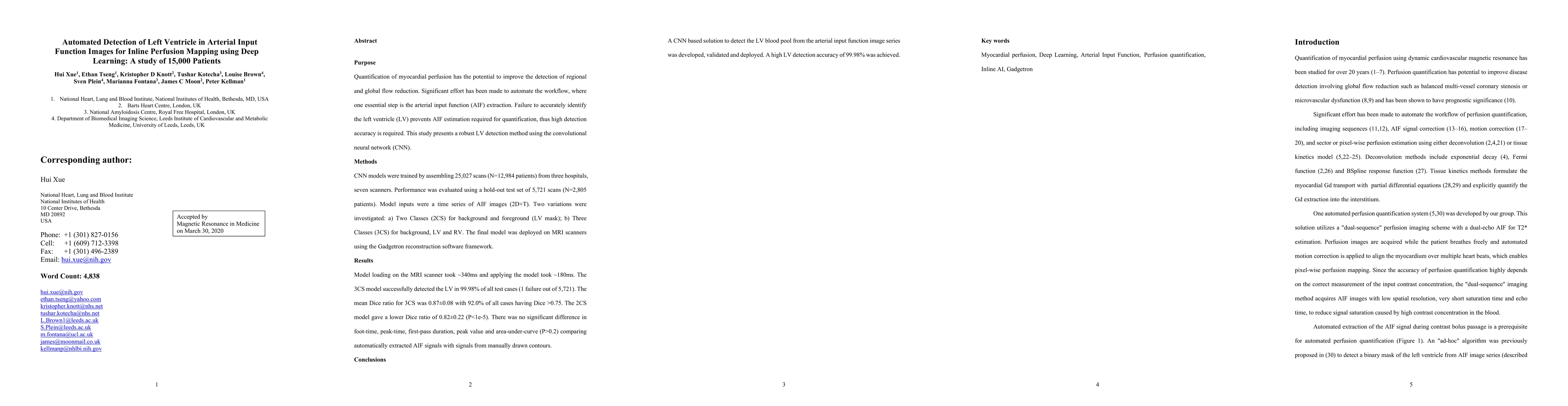
Quantification of myocardial perfusion has the potential to improve detection of regional and global flow reduction. Significant effort has been made to automate the workflow, where one essential step is the arterial input function (AIF) extraction. Since failure here invalidates quantification, high accuracy is required. For this purpose, this study presents a robust AIF detection method using the convolutional neural net (CNN) model. CNN models were trained by assembling 25,027 scans (N=12,984 patients) from three hospitals, seven scanners. A test set of 5,721 scans (N=2,805 patients) evaluated model performance. The 2D+T AIF time series was inputted into CNN. Two variations were investigated: a) Two Classes (2CS) for background and foreground (LV mask); b) Three Classes (3CS) for background, foreground LV and RV. Final model was deployed on MR scanners via the Gadgetron InlineAI. Model loading time on MR scanner was ~340ms and applying it took ~180ms. The 3CS model successfully detect LV for 99.98% of all test cases (1 failed out of 5,721 cases). The mean Dice ratio for 3CS was 0.87+/-0.08 with 92.0% of all test cases having Dice ratio >0.75, while the 2CS model gave lower Dice of 0.82+/-0.22 (P<1e-5). Extracted AIF signals using CNN were further compared to manual ground-truth for foot-time, peak-time, first-pass duration, peak value and area-under-curve. No significant differences were found for all features (P>0.2). This study proposed, validated, and deployed a robust CNN solution to detect the LV for the extraction of the AIF signal used in fully automated perfusion flow mapping. A very large data cohort was assembled and resulting models were deployed to MR scanners for fully inline AI in clinical hospitals.

AI Key Findings

Get AI-generated insights about this paper's methodology, results, and significance.

Paper Details

PDF Preview

Download PDF

Key Terms

cnn (0.276)
deployed (0.231)
quantification (0.203)
patients (0.186)
extraction (0.164)
test (0.157)
detect (0.154)
peak (0.152)
cases (0.149)
classes (0.147)
flow (0.135)
background (0.135)
fully (0.135)
robust (0.135)
ratio (0.128)
detection (0.124)
model performance (0.122)
seven (0.118)
significant (0.116)
high accuracy (0.116)

Citation Network

Current paper (gray), citations (green), references (blue)

Current Paper
Citations
References
Click to view
Title Authors Year Actions
The Role of Artificial Intelligence in the Prediction, Diagnosis, and Management of Cardiovascular Diseases: A Narrative Review
Mohammed Farooque W Shaikh, Murtaza S Mama, Sri Harika Proddaturi, Juan Vidal, Pritika Gnanasekaran, Mekala S Kumar, Cleve J Clarke, K. S. Reddy, H. M. Bello, Naama Raquib, Zoya Morani
2025
View Paper
Scanner-based real-time three-dimensional brain + body slice-to-volume reconstruction for T2-weighted 0.55-T low-field fetal magnetic resonance imaging
A. Uus, S. Neves Silva, J. Aviles Verdera, K. Payette, Megan Hall, K. Colford, Aysha Luis, H. Sousa, Z. Ning, T. Roberts, S. McElroy, M. Deprez, J. Hajnal, M. Rutherford, Lisa Story, J. Hutter
2025
View Paper
Prognostic value of novel cardiovascular magnetic resonance transit times beyond the pulmonary circulation in patients with ventricular dysfunction.
Teresa Sevilla, C. Baladrón, M. de Miguel-Álava, G. Rojas-Lavado, E. Gonzalez-Bartol, A. Revilla-Orodea, C. Aristizábal-Duque, M. Carrasco-Moraleja, Miguel Fernández-Garrote, J. S. San Román
2024
View Paper
Pulmonary transit time is a predictor of outcomes in heart failure: a cardiovascular magnetic resonance first-pass perfusion study
Jonathan Farley, L. Brown, P. Garg, A. Wahab, Joel R. L. Klassen, N. Jex, S. Thirunavukarasu, A. Chowdhary, N. Sharrack, M. Gorecka, Hui Xue, Nigel Artis, E. Levelt, E. Dall’Armellina, Peter Kellman, John P. Greenwood, S. Plein, P. Swoboda
2024
View Paper
The role of artificial intelligence in cardiovascular magnetic resonance imaging.
Afolasayo A. Aromiwura, João L. Cavalcante, Raymond Y Kwong, Aryan Ghazipour, Amir Amini, Jeroen J. Bax, Subha V. Raman, G. Pontone, Dinesh K Kalra
2024
View Paper
Scanner-based real-time 3D brain+body slice-to-volume reconstruction for T2-weighted 0.55T low field fetal MRI
A. Uus, S. Neves Silva, J. Aviles Verdera, K. Payette, M. Hall, K. Colford, A. Luis, H. Sousa, Z. Ning, T. Roberts, S. McElroy, M. Deprez, J. Hajnal, M. Rutherford, L. Story, J. Hutter
2024
View Paper
A simplified method to correct saturation of arterial input function for cardiac magnetic resonance first-pass perfusion imaging: validation with simultaneously acquired PET
Ran Li, Masoud Edalati, D. Muccigrosso, Jeffrey M. C. Lau, R. Laforest, P. Woodard, Jie Zheng
2023
View Paper
Approach to Imaging Ischemia in Women.
P. Parwani, Adeba Mohammad, Y. Liberman, D. Litmanovich
2023
View Paper
Stress native T1 and native T2 mapping compared to myocardial perfusion reserve in long-term follow-up of severe Covid-19
J. Nickander, R. Steffen Johansson, K. Lodin, Anton Wahrby, D. Loewenstein, J. Bruchfeld, M. Runold, H. Xue, P. Kellman, H. Engblom
2023
View Paper
Machine Learning Approaches in Diagnosis, Prognosis and Treatment Selection of Cardiac Amyloidosis
A. Allegra, Giuseppe Mirabile, A. Tonacci, Sarah Genovese, G. Pioggia, S. Gangemi
2023
View Paper
Left Ventricle Detection from Cardiac Magnetic Resonance Relaxometry Images Using Visual Transformer
Lisa Anita De Santi, A. Meloni, M. Santarelli, L. Pistoia, A. Spasiano, T. Casini, M. Putti, L. Cuccia, Filippo Cademartiri, V. Positano
2023
View Paper
An Overview of Deep Learning Methods for Left Ventricle Segmentation
M. Shoaib, Joon Huang Chuah, Raza Ali, K. Hasikin, A. Khalil, Y. Hum, Y. Tee, S. Dhanalakshmi, K. Lai
2023
View Paper
Sex- and age-specific normal values for automated quantitative pixel-wise myocardial perfusion cardiovascular magnetic resonance
L. Brown, G. Gulsin, S. Onciul, D. Broadbent, J. Yeo, A. Wood, C. Saunderson, Arka Das, N. Jex, A. Chowdhary, S. Thirunavukarasu, N. Sharrack, K. Knott, E. Levelt, P. Swoboda, H. Xue, J. Greenwood, J. Moon, D. Adlam, G. Mccann, P. Kellman, S. Plein
2022
View Paper
State-of-the-Art of Myocardial Perfusion by CMR: A Practical View
G. Pons-Lladó, P. Kellman
2022
View Paper
Editorial for “Impact of Temporal Resolution and Methods for Correction on Cardiovascular Magnetic Resonance Perfusion Quantification”
S. Rapacchi, M. Viallon, P. Croisille
2022
View Paper
Artificial Intelligence in Cardiology—A Narrative Review of Current Status
G. Koulaouzidis, T. Jadczyk, D. Iakovidis, Anastasios Koulaouzidis, Marc Bisnaire, D. Charisopoulou
2022
View Paper
Free‐breathing motion‐informed locally low‐rank quantitative 3D myocardial perfusion imaging
Tobias Hoh, V. Vishnevskiy, M. Polacin, R. Manka, M. Fuetterer, S. Kozerke
2022
View Paper
Imaging gravity-induced lung water redistribution with automated inline processing at 0.55 T cardiovascular magnetic resonance
F. Seemann, A. Javed, Rachel Chae, R. Ramasawmy, K. O’Brien, S. Baute, H. Xue, R. Lederman, A. Campbell-Washburn
2022
View Paper
Cardiac MR: From Theory to Practice
T. Ismail, W. Strugnell, C. Coletti, Maša Božić-Iven, Sebastian Weingärtner, K. Hammernik, T. Correia, T. Küstner
2022
View Paper
Ultrasonic Omics Based on Intelligent Classification Algorithm in Hormone Receptor Expression and Efficacy Evaluation of Breast Cancer
Musu Ala, Jianlin Wu
2022
View Paper
Deep learning applications in myocardial perfusion imaging, a systematic review and meta-analysis
E. Alskaf, Utkarsh Dutta, C. Scannell, A. Chiribiri
2022
View Paper
Echocardiographic Features of Patients with Coronary Heart Disease and Angina Pectoris under Deep Learning Algorithms
Xianjing Han, G. Liang
2021
View Paper
A Patch-Wise Deep Learning Approach for Myocardial Blood Flow Quantification with Robustness to Noise and Nonrigid Motion
K. Youssef, B. Heydari, Luis Zamudio Rivero, Taylor Beaulieu, K. Cheema, R. Dharmakumar, B. Sharif
2021
View Paper
Deep Learning-Based Segmentation and Uncertainty Assessment for Automated Analysis of Myocardial Perfusion MRI Datasets Using Patch-Level Training and Advanced Data Augmentation
D. Yalcinkaya, K. Youssef, B. Heydari, Luis Zamudio, R. Dharmakumar, B. Sharif
2021
View Paper
A pilot randomised controlled trial of a structured, home-based exercise programme on cardiovascular structure and function in kidney transplant recipients: the ECSERT study design and methods
R. Billany, Noemi Vadaszy, N. Bishop, T. Wilkinson, Sherna F. Adenwalla, Kate A Robinson, Kathryn Croker, E. Brady, J. Wormleighton, K. Parke, N. Cooper, A. Webster, J. Barratt, G. Mccann, J. Burton, Alice C. Smith, M. Graham-Brown
2021
View Paper
Automated Quantitative Stress Perfusion Cardiac Magnetic Resonance in Pediatric Patients
C. Scannell, Hadeer Hasaneen, G. Greil, T. Hussain, R. Razavi, Jack Lee, K. Pushparajah, Phuoc Duong, A. Chiribiri
2021
View Paper
Emerging Techniques in Cardiac Magnetic Resonance Imaging
Rui Guo, Sebastian Weingärtner, Paulina Šiurytė, Christian T Stoeck, Maximilian Füetterer, Adrienne E Campbell-Washburn, Avan Suinesiaputra, M. Jerosch-Herold, R. Nezafat
2021
View Paper
Myocardial Perfusion Defects in Hypertrophic Cardiomyopathy Mutation Carriers
R. Hughes, C. Camaioni, J. Augusto, K. Knott, Ellie Quinn, G. Captur, A. Seraphim, G. Joy, P. Syrris, P. Elliott, Saidi A Mohiddin, P. Kellman, H. Xue, L. Lopes, J. Moon
2021
View Paper
Landmark Detection in Cardiac MRI by Using a Convolutional Neural Network
H. Xue, J. Artico, M. Fontana, J. Moon, R. Davies, P. Kellman
2021
View Paper
The cardiac surgeon's guide to artificial intelligence
R. Nedadur, Bo Wang, B. Yanagawa
2021
View Paper
Use of quantitative cardiovascular magnetic resonance myocardial perfusion mapping for characterization of ischemia in patients with left internal mammary coronary artery bypass grafts
A. Seraphim, K. Knott, A. Beirne, J. Augusto, K. Menacho, J. Artico, G. Joy, R. Hughes, A. Bhuva, R. Torii, H. Xue, T. Treibel, R. Davies, J. Moon, Daniel A. Jones, P. Kellman, C. Manisty
2021
View Paper
Prognostic Value of Pulmonary Transit Time and Pulmonary Blood Volume Estimation Using Myocardial Perfusion CMR
A. Seraphim, K. Knott, K. Menacho, J. Augusto, R. Davies, I. Pierce, G. Joy, A. Bhuva, H. Xue, T. Treibel, J. Cooper, Steffen Erhard Petersen, M. Fontana, A. Hughes, J. Moon, C. Manisty, P. Kellman
2021
View Paper
Influence of motion correction on the visual analysis of cardiac magnetic resonance stress perfusion imaging
F. von Knobelsdorff-Brenkenhoff, Stephanie Reiter, A. Menini, M. Janich, T. Schunke, Karl Ziegler, R. Scheck, B. Höfling, G. Pilz
2021
View Paper
A comparison of standard and high dose adenosine protocols in routine vasodilator stress cardiovascular magnetic resonance: dosage affects hyperaemic myocardial blood flow in patients with severe left ventricular systolic impairment
L. Brown, C. Saunderson, Arka Das, Thomas P. Craven, E. Levelt, K. Knott, E. Dall’Armellina, H. Xue, J. Moon, J. Greenwood, P. Kellman, P. Swoboda, S. Plein
2021
View Paper
The Higher You Climb, the Better the View: Quantitative CMR Perfusion Mapping for CAD.
R. Kwong, Y. Chandrashekhar
2020
View Paper
Quantitative perfusion-CMR is significantly influenced by the placement of the arterial input function
Ibnul Mia, Melanie Le, C. Arendt, D. Brand, Sina Bremekamp, Tommaso D’Angelo, V. Puntmann, E. Nagel
2020
View Paper
Artificial intelligence and cardiovascular imaging: A win–win combination
L. Badano, Daria M. Keller, D. Muraru, C. Torlasco, G. Parati
2020
View Paper
Landmark detection in Cardiac Magnetic Resonance Imaging Using A Convolutional Neural Network
H. Xue, J. Artico, M. Fontana, J. Moon, R. Davies, P. Kellman
2020
View Paper
Feasibility of free-breathing quantitative myocardial perfusion using multi-echo Dixon magnetic resonance imaging
C. Scannell, T. Correia, A. Villa, T. Schneider, Jack Lee, M. Breeuwer, A. Chiribiri, M. Henningsson
2020
View Paper
Pathophysiology, classification, and MRI parallels in microvascular disease of the heart and brain
M. A. Thomas, Saman Hazany, B. Ellingson, P. Hu, Kim-Lien Nguyen
2020
View Paper
Females have higher myocardial perfusion, blood volume and extracellular volume compared to males – an adenosine stress cardiovascular magnetic resonance study
J. Nickander, R. Themudo, A. Sigfridsson, H. Xue, P. Kellman, M. Ugander
2020
View Paper
Influence of contrast agent dispersion on bolus‐based MRI myocardial perfusion measurements: A computational fluid dynamics study
Johannes Martens, S. Panzer, J. V. D. van den Wijngaard, M. Siebes, L. Schreiber
2019
View Paper
Automated Inline Analysis of Myocardial Perfusion MRI with Deep Learning
H. Xue, R. Davies, L. Brown, K. Knott, T. Kotecha, M. Fontana, S. Plein, J. Moon, P. Kellman
2019
View Paper
Hypertrophic cardiomyopathy deserves better – ditch the 16 segments
R. Hughes, J. Moon, G. Captur
2019
View Paper

Comments (0)

This website uses cookies to ensure you get the best experience. Learn more