Automated detection of lung nodules in low-dose computed tomography
Publication
Metrics
AI Quick Summary
This paper presents a CAD system for detecting pulmonary nodules in low-dose CT scans, developed within the MAGIC-5 project. The system employs a 3D dot-enhancement filter and a neural classifier, achieving high sensitivity (85%) and low false positives (1-9 FP/scan).
Paper Preview
Abstract
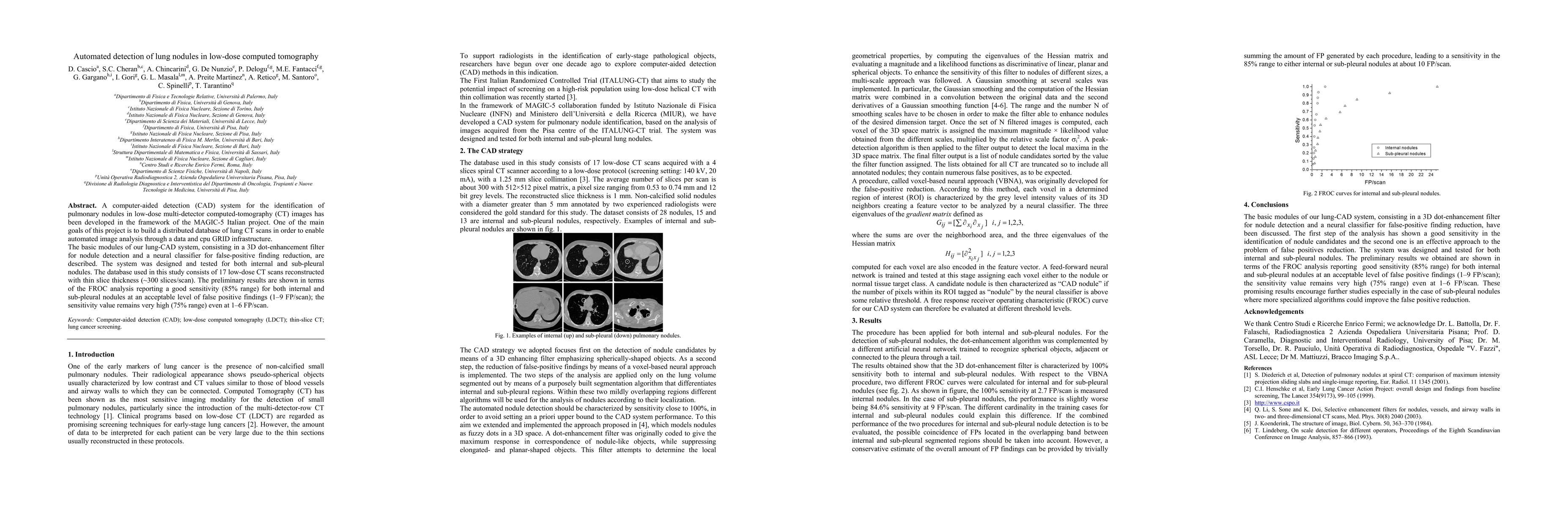
A computer-aided detection (CAD) system for the identification of pulmonary nodules in low-dose multi-detector computed-tomography (CT) images has been developed in the framework of the MAGIC-5 Italian project. One of the main goals of this project is to build a distributed database of lung CT scans in order to enable automated image analysis through a data and cpu GRID infrastructure. The basic modules of our lung-CAD system, consisting in a 3D dot-enhancement filter for nodule detection and a neural classifier for false-positive finding reduction, are described. The system was designed and tested for both internal and sub-pleural nodules. The database used in this study consists of 17 low-dose CT scans reconstructed with thin slice thickness (~300 slices/scan). The preliminary results are shown in terms of the FROC analysis reporting a good sensitivity (85% range) for both internal and sub-pleural nodules at an acceptable level of false positive findings (1-9 FP/scan); the sensitivity value remains very high (75% range) even at 1-6 FP/scan
AI Key Findings
Get AI-generated insights about this paper's methodology, results, significance, and more — seven facets brought into focus.
Impact
Paper Details
PDF Preview
Key Terms
Citation Network
Current paper (gray), citations (green), references (blue)
Display is limited for performance on very large graphs.
Discussion 0