Automated detection of motion artifacts in brain MR images using deep learning and explainable artificial intelligence
Publication
Metrics
AI Quick Summary
This study presents a deep learning model using 2D CNNs to automatically detect motion artifacts in T1-weighted brain MRI images, achieving 85% precision and 80% recall. Grad-CAM heatmaps provide interpretability, aiding in the identification of failure modes and enhancing model reliability.
Paper Preview
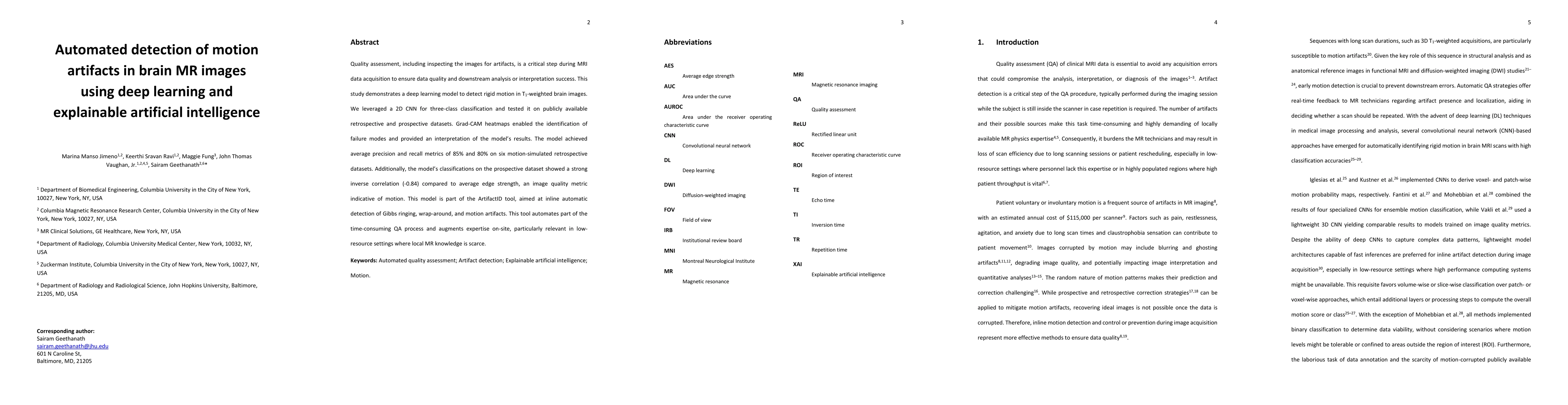
Abstract
Quality assessment, including inspecting the images for artifacts, is a critical step during MRI data acquisition to ensure data quality and downstream analysis or interpretation success. This study demonstrates a deep learning model to detect rigid motion in T1-weighted brain images. We leveraged a 2D CNN for three-class classification and tested it on publicly available retrospective and prospective datasets. Grad-CAM heatmaps enabled the identification of failure modes and provided an interpretation of the model's results. The model achieved average precision and recall metrics of 85% and 80% on six motion-simulated retrospective datasets. Additionally, the model's classifications on the prospective dataset showed a strong inverse correlation (-0.84) compared to average edge strength, an image quality metric indicative of motion. This model is part of the ArtifactID tool, aimed at inline automatic detection of Gibbs ringing, wrap-around, and motion artifacts. This tool automates part of the time-consuming QA process and augments expertise on-site, particularly relevant in low-resource settings where local MR knowledge is scarce.
AI Key Findings
Get AI-generated insights about this paper's methodology, results, significance, and more — seven facets brought into focus.
Impact
Paper Details
Authors
PDF Preview
Key Terms
Citation Network
Current paper (gray), citations (green), references (blue)
Display is limited for performance on very large graphs.
Discussion 0