Automated Diagnosis of Lymphoma with Digital Pathology Images Using Deep Learning
Publication
Metrics
AI Quick Summary
This study developed a deep learning convolutional neural network model to diagnose four types of lymphoma from digital pathology images, achieving 95% accuracy for individual image predictions and 10% accuracy for set-based predictions. The preliminary results suggest potential for enhancing pathologist productivity through automated diagnostic screening.
Paper Preview
Abstract
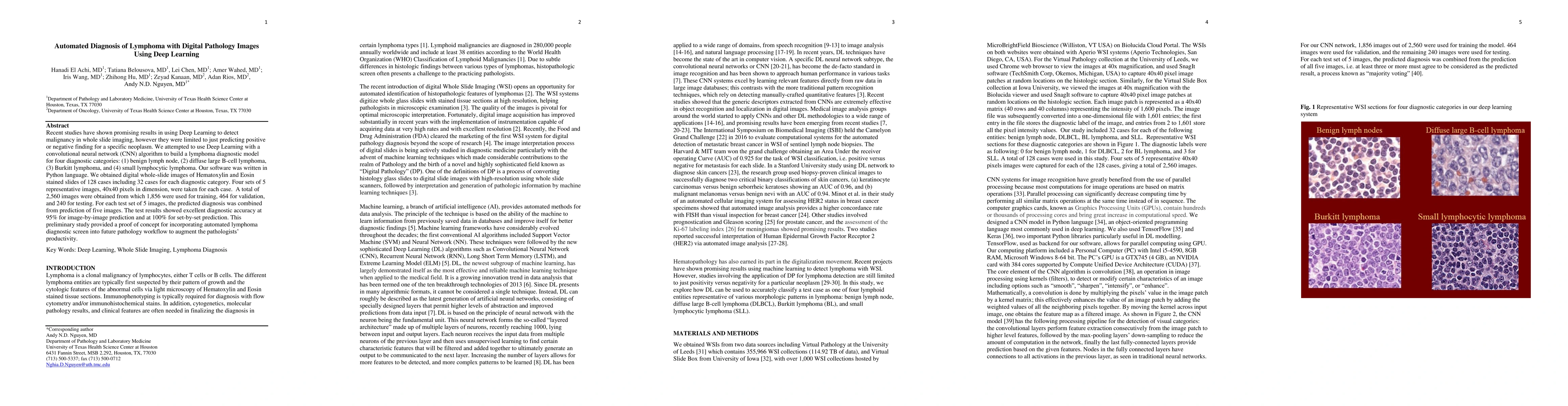
Recent studies have shown promising results in using Deep Learning to detect malignancy in whole slide imaging. However, they were limited to just predicting positive or negative finding for a specific neoplasm. We attempted to use Deep Learning with a convolutional neural network algorithm to build a lymphoma diagnostic model for four diagnostic categories: benign lymph node, diffuse large B cell lymphoma, Burkitt lymphoma, and small lymphocytic lymphoma. Our software was written in Python language. We obtained digital whole slide images of Hematoxylin and Eosin stained slides of 128 cases including 32 cases for each diagnostic category. Four sets of 5 representative images, 40x40 pixels in dimension, were taken for each case. A total of 2,560 images were obtained from which 1,856 were used for training, 464 for validation and 240 for testing. For each test set of 5 images, the predicted diagnosis was combined from prediction of 5 images. The test results showed excellent diagnostic accuracy at 95% for image-by-image prediction and at 10% for set-by-set prediction. This preliminary study provided a proof of concept for incorporating automated lymphoma diagnostic screen into future pathology workflow to augment the pathologists' productivity.
AI Key Findings
Get AI-generated insights about this paper's methodology, results, significance, and more — seven facets brought into focus.
Impact
Paper Details
PDF Preview
Key Terms
Citation Network
Current paper (gray), citations (green), references (blue)
Display is limited for performance on very large graphs.
Discussion 0