Publication
Metrics
AI Quick Summary
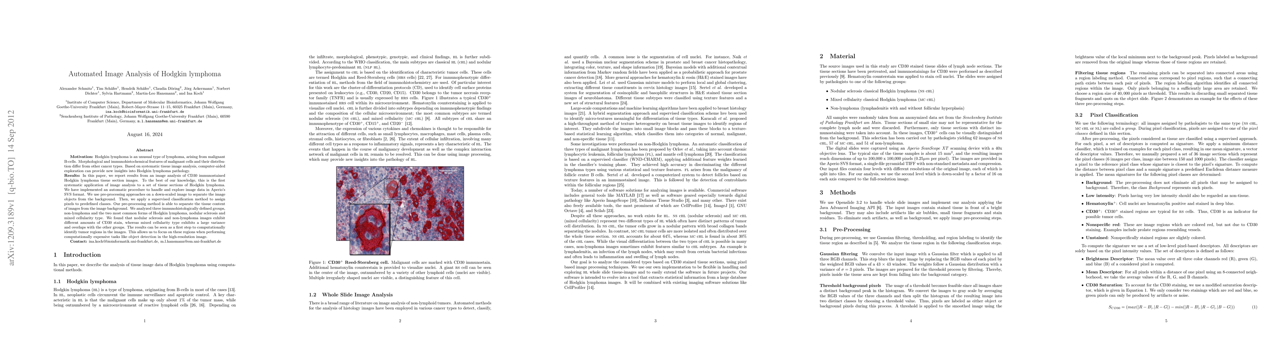
This paper presents an automated image analysis method for CD30 immunostained Hodgkin lymphoma tissue sections, marking the first systematic application of such analysis. The study uses pre-processing and supervised classification to identify tumor regions, revealing distinct CD30 staining patterns among nodular sclerosis, mixed cellularity types, and non-lymphoma controls.
Paper Preview
Abstract
Hodgkin lymphoma is an unusual type of lymphoma, arising from malignant B-cells. Morphological and immunohistochemical features of malignant cells and their distribution differ from other cancer types. Based on systematic tissue image analysis, computer-aided exploration can provide new insights into Hodgkin lymphoma pathology. In this paper, we report results from an image analysis of CD30 immunostained Hodgkin lymphoma tissue section images. To the best of our knowledge, this is the first systematic application of image analysis to a set of tissue sections of Hodgkin lymphoma. We have implemented an automatic procedure to handle and explore image data in Aperio's SVS format. We use pre-processing approaches on a down-scaled image to separate the image objects from the background. Then, we apply a supervised classification method to assign pixels to predefined classes. Our pre-processing method is able to separate the tissue content of images from the image background. We analyzed three immunohistologically defined groups, non-lymphoma and the two most common forms of Hodgkin lymphoma, nodular sclerosis and mixed cellularity type. We found that nodular sclerosis and non-lymphoma images exhibit different amounts of CD30 stain, whereas mixed cellularity type exhibits a large variance and overlaps with the other groups. The results can be seen as a first step to computationally identify tumor regions in the images. This allows us to focus on these regions when performing computationally expensive tasks like object detection in the high-resolution image.
AI Key Findings
Get AI-generated insights about this paper's methodology, results, significance, and more — seven facets brought into focus.
Impact
Paper Details
PDF Preview
Key Terms
Citation Network
Current paper (gray), citations (green), references (blue)
Display is limited for performance on very large graphs.
Discussion 0