Automated Multi-Channel Segmentation for the 4D Myocardial Velocity Mapping Cardiac MR
Publication
Metrics
AI Quick Summary
This paper proposes an automated deep learning framework to enhance the segmentation of myocardial contours in 4D myocardial velocity mapping cardiac MR images using multi-channel data fusion. The method shows improved performance over standard U-Net based segmentations, as evidenced by higher Dice scores and more accurate quantification of myocardial velocities.
Paper Preview
Abstract
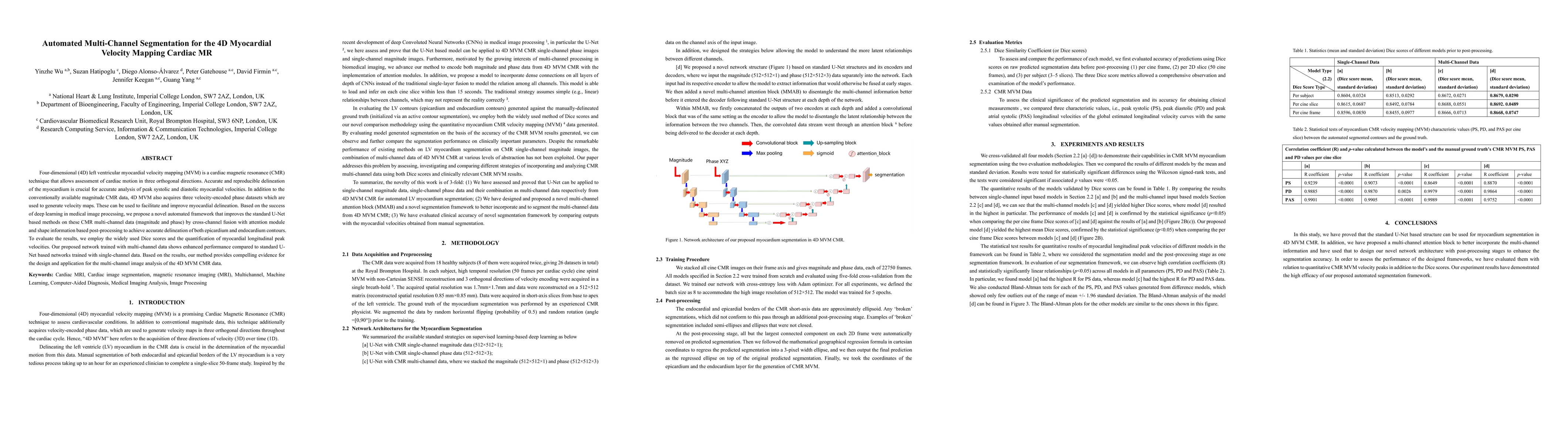
Four-dimensional (4D) left ventricular myocardial velocity mapping (MVM) is a cardiac magnetic resonance (CMR) technique that allows assessment of cardiac motion in three orthogonal directions. Accurate and reproducible delineation of the myocardium is crucial for accurate analysis of peak systolic and diastolic myocardial velocities. In addition to the conventionally available magnitude CMR data, 4D MVM also acquires three velocity-encoded phase datasets which are used to generate velocity maps. These can be used to facilitate and improve myocardial delineation. Based on the success of deep learning in medical image processing, we propose a novel automated framework that improves the standard U-Net based methods on these CMR multi-channel data (magnitude and phase) by cross-channel fusion with attention module and shape information based post-processing to achieve accurate delineation of both epicardium and endocardium contours. To evaluate the results, we employ the widely used Dice scores and the quantification of myocardial longitudinal peak velocities. Our proposed network trained with multi-channel data shows enhanced performance compared to standard U-Net based networks trained with single-channel data. Based on the results, our method provides compelling evidence for the design and application for the multi-channel image analysis of the 4D MVM CMR data.
AI Key Findings
Get AI-generated insights about this paper's methodology, results, significance, and more — seven facets brought into focus.
Impact
Paper Details
Authors
PDF Preview
Key Terms
Citation Network
Current paper (gray), citations (green), references (blue)
Display is limited for performance on very large graphs.
Discussion 0