Automated Real-Space Lattice Extraction for Atomic Force Microscopy Images
Publication
Metrics
AI Quick Summary
This research paper introduces AiSurf, a free tool for automated analysis of atomically resolved AFM images using SIFT and clustering algorithms, enabling extraction of lattice vectors and unit cells with minimal user input, thereby significantly reducing the time and expertise required for interpreting complex AFM images. The method demonstrated robustness across various surfaces, enhancing the efficiency of scanning probe microscopy image analysis.
Paper Preview
Abstract
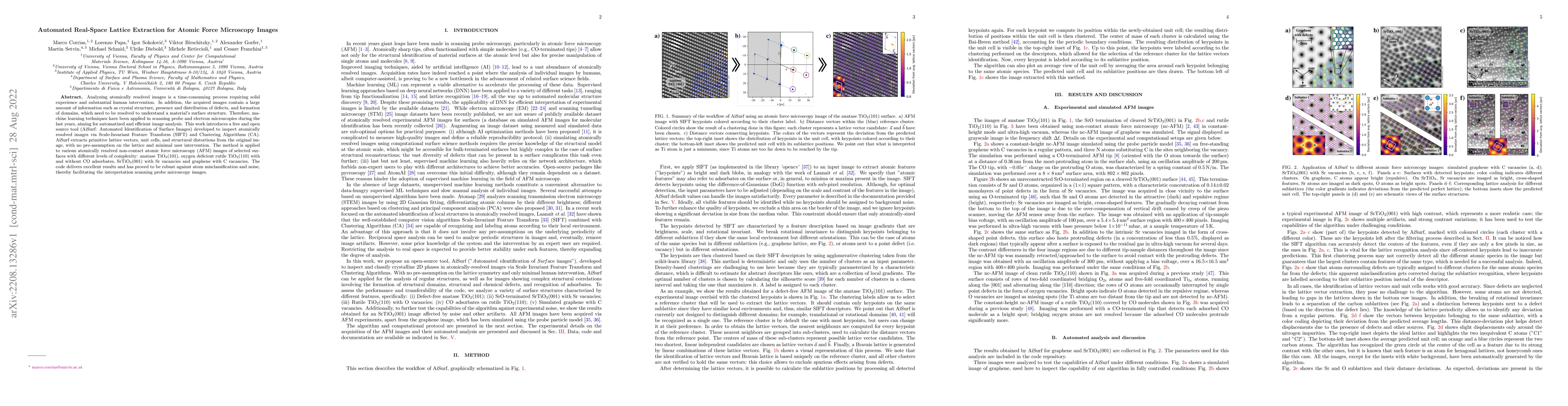
Analyzing atomically resolved images is a time-consuming process requiring solid experience and substantial human intervention. In addition, the acquired images contain a large amount of information such as crystal structure, presence and distribution of defects, and formation of domains, which need to be resolved to understand a material's surface structure. Therefore, machine learning techniques have been applied in scanning probe and electron microscopies during the last years, aiming for automatized and efficient image analysis. This work introduces a free and open source tool (AiSurf: Automated Identification of Surface Images) developed to inspect atomically resolved images via Scale-Invariant Feature Transform (SIFT) and Clustering Algorithms (CA). AiSurf extracts primitive lattice vectors, unit cells, and structural distortions from the original image, with no pre-assumption on the lattice and minimal user intervention. The method is applied to various atomically resolved non-contact atomic force microscopy (AFM) images of selected surfaces with different levels of complexity: anatase TiO2(101), oxygen deficient rutile TiO2(110) with and without CO adsorbates, SrTiO3(001) with Sr vacancies and graphene with C vacancies. The code delivers excellent results and has proved to be robust against atom misclassification and noise, thereby facilitating the interpretation scanning probe microscopy images.
AI Key Findings
Get AI-generated insights about this paper's methodology, results, significance, and more — seven facets brought into focus.
Impact
Paper Details
Authors
PDF Preview
Key Terms
Citation Network
Current paper (gray), citations (green), references (blue)
Display is limited for performance on very large graphs.
Discussion 0