Summary
Subarachnoid hemorrhage (SAH), typically due to intracranial aneurysms, demands precise imaging for effective treatment. Digital Subtraction Angiography (DSA), despite being the gold standard, broadly visualizes cerebral blood flow, potentially masking key details in areas. This study introduces an approach integrating a 3D vascular atlas with 2D DSA images to allow targeted quantitative analysis in these crucial regions, thus enhancing diagnostic accuracy during interventions. Initially, DSA data was examined to ascertain the injection site. Following this, the appropriate viewing angle was determined to align accurately with the 3D vascular atlas. Utilizing this atlas, regions corresponding to the areas indicated as perfused were selected. Concurrently, a mask representing the perfused areas was created from the DSA sequence. This mask facilitated the initial coarse alignment of the projected 3D atlas to the DSA perfused territory deformable registration techniques, ensuring a precise overlay with the DSAs perfused territories. The performance of each overlay was measured using the Structural Similarity Index Measure (SSIM). The coregistration process revealed that deformable registrations was essential to achieve precise overlays of the 3D atlas projections with the 2D DSA perfused areas. This approach enabled the extraction of targeted quantitative angiography parameters, essential for detailed vascular assessment in subarachnoid hemorrhage cases. The integration of 3D atlas registration with 2D DSA projections facilitates a more precise and targeted diagnostic process for SAH during critical interventions. This image processing strategy enhances the visualization of affected arterial territories, potentially improving the accuracy of diagnostics and supporting better informed clinical decisions at the time of intervention
AI Key Findings
Generated Sep 02, 2025
Methodology
The study integrates a 3D vascular atlas with 2D DSA projections for targeted quantitative angiography analysis in SAH cases. It involves manual labeling of DSA sequences, cone beam projection of 3D atlas regions, creation of DSA masks, affine and B-spline registrations, and evaluation using SSIM.
Key Results
- The approach enabled precise localization and detailed assessment of blood flow dynamics within specific vascular territories.
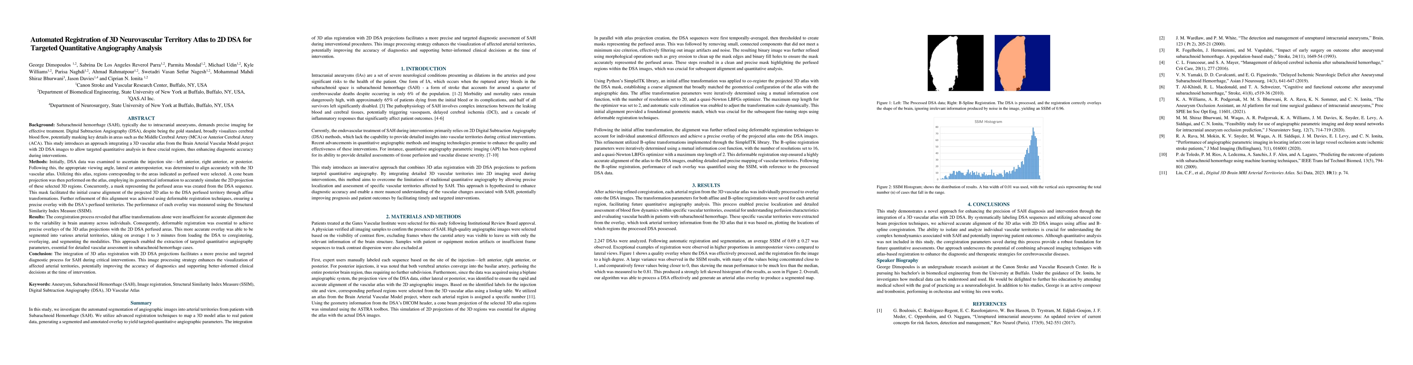
- An average SSIM of 0.69 ± 0.27 was observed across 2,247 DSAs, with higher proportions of exceptional registration in anteroposterior views.
- The method facilitated enhanced visualization of affected arterial territories, potentially improving diagnostic accuracy and clinical decision-making.
Significance
This research is significant as it addresses the limitations of traditional quantitative angiography by allowing precise localization and assessment of specific vascular territories affected by SAH, potentially improving prognosis and patient outcomes.
Technical Contribution
The main technical contribution is the development of an image processing strategy that combines 3D atlas registration with 2D DSA projections for precise and targeted diagnostic assessment of SAH during interventional procedures.
Novelty
This work stands out by introducing a novel approach that integrates 3D vascular atlas registration with 2D DSA projections, overcoming the limitations of traditional quantitative angiography methods and enhancing diagnostic precision for SAH cases.
Limitations
- The study did not include quantitative analysis, focusing mainly on the registration process.
- The performance evaluation relied on SSIM, which might not capture all aspects of diagnostic accuracy.
Future Work
- Future work could involve incorporating quantitative analysis to evaluate vascular health based on the extracted parameters.
- Exploration of alternative registration techniques or optimization methods for improved alignment accuracy.
Paper Details
PDF Preview
Citation Network
Current paper (gray), citations (green), references (blue)
Display is limited for performance on very large graphs.
Similar Papers
Found 4 papers3D/2D Registration of Angiograms using Silhouette-based Differentiable Rendering
Nazim Haouchine, Sarah Frisken, Taewoong Lee
In-Silico Investigation of 3D Quantitative Angiography for Internal Carotid Aneurysms Using Biplane Imaging and 3D Vascular Geometry Constraints
Kyle A. Williams, Swetadri Vasan Setlur Nagesh, Ciprian N. Ionita et al.
No citations found for this paper.
Comments (0)