Automated robotic intraoperative ultrasound for brain surgery
Publication
Metrics
AI Quick Summary
This paper introduces a robotic framework for automated intraoperative ultrasound (iUS) during brain tumour resection, aiming to simplify anatomy localisation and data interpretation, which has been a challenge for surgeons. The framework was tested on a soft-tissue-mimicking brain phantom to simulate real-time iUS scanning.
Paper Preview
Abstract
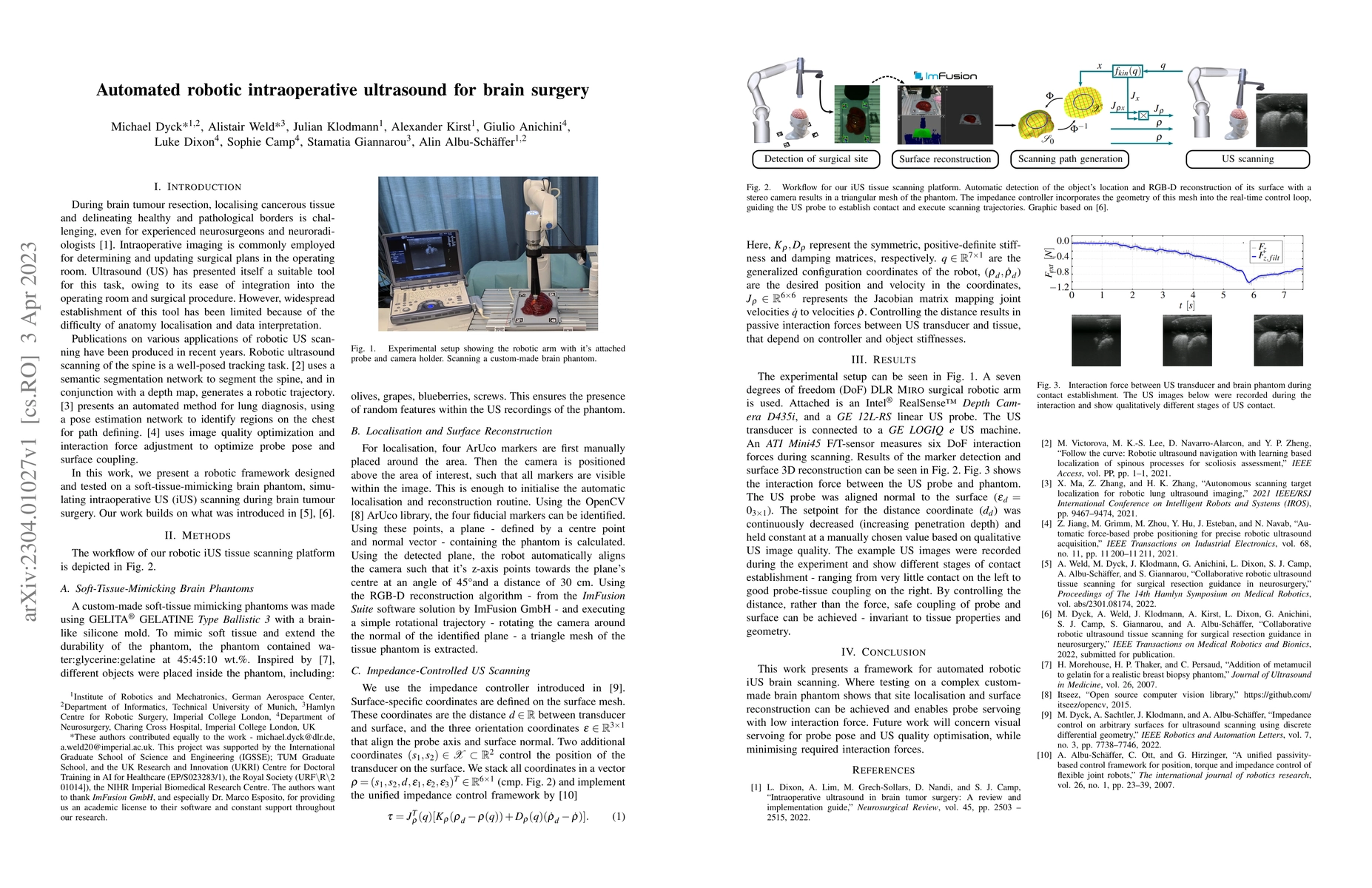
During brain tumour resection, localising cancerous tissue and delineating healthy and pathological borders is challenging, even for experienced neurosurgeons and neuroradiologists. Intraoperative imaging is commonly employed for determining and updating surgical plans in the operating room. Ultrasound (US) has presented itself a suitable tool for this task, owing to its ease of integration into the operating room and surgical procedure. However, widespread establishment of this tool has been limited because of the difficulty of anatomy localisation and data interpretation. In this work, we present a robotic framework designed and tested on a soft-tissue-mimicking brain phantom, simulating intraoperative US (iUS) scanning during brain tumour surgery.
AI Key Findings
Get AI-generated insights about this paper's methodology, results, significance, and more — seven facets brought into focus.
Impact
Paper Details
Authors
PDF Preview
Key Terms
Citation Network
Current paper (gray), citations (green), references (blue)
Display is limited for performance on very large graphs.
Discussion 0