Summary
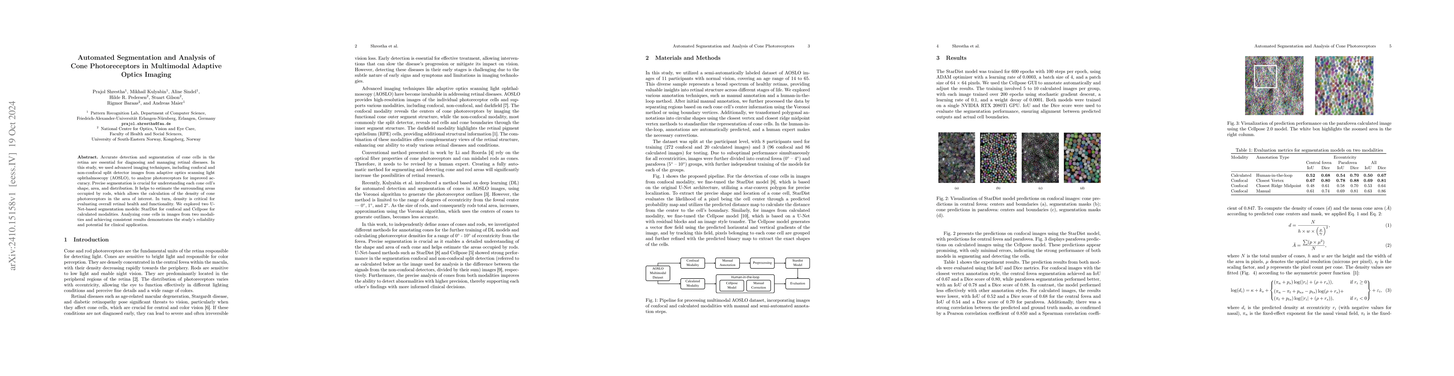
Accurate detection and segmentation of cone cells in the retina are essential for diagnosing and managing retinal diseases. In this study, we used advanced imaging techniques, including confocal and non-confocal split detector images from adaptive optics scanning light ophthalmoscopy (AOSLO), to analyze photoreceptors for improved accuracy. Precise segmentation is crucial for understanding each cone cell's shape, area, and distribution. It helps to estimate the surrounding areas occupied by rods, which allows the calculation of the density of cone photoreceptors in the area of interest. In turn, density is critical for evaluating overall retinal health and functionality. We explored two U-Net-based segmentation models: StarDist for confocal and Cellpose for calculated modalities. Analyzing cone cells in images from two modalities and achieving consistent results demonstrates the study's reliability and potential for clinical application.
AI Key Findings
Get AI-generated insights about this paper's methodology, results, and significance.
Paper Details
PDF Preview
Citation Network
Current paper (gray), citations (green), references (blue)
Display is limited for performance on very large graphs.
Similar Papers
Found 4 papersGeneralist Segmentation Algorithm for Photoreceptors Analysis in Adaptive Optics Imaging
Andreas Maier, Mikhail Kulyabin, Aline Sindel et al.
Adaptive Interactive Segmentation for Multimodal Medical Imaging via Selection Engine
Zhi Li, Shuai Wang, Yaqi Wang et al.
No citations found for this paper.
Comments (0)