Quick Summary:

This paper proposes a deep learning model, Retinal Fluid Segmentation Network (ReF-Net), to accurately segment retinal fluid volumes from optical coherence tomography (OCT) and OCT angiography (OCTA) scans. The model shows high accuracy and robustness, with improved performance when using combined OCTA and OCT data, suggesting volumetric retinal fluid analysis provides more comprehensive diagnostic information than 2D projections.

Summary

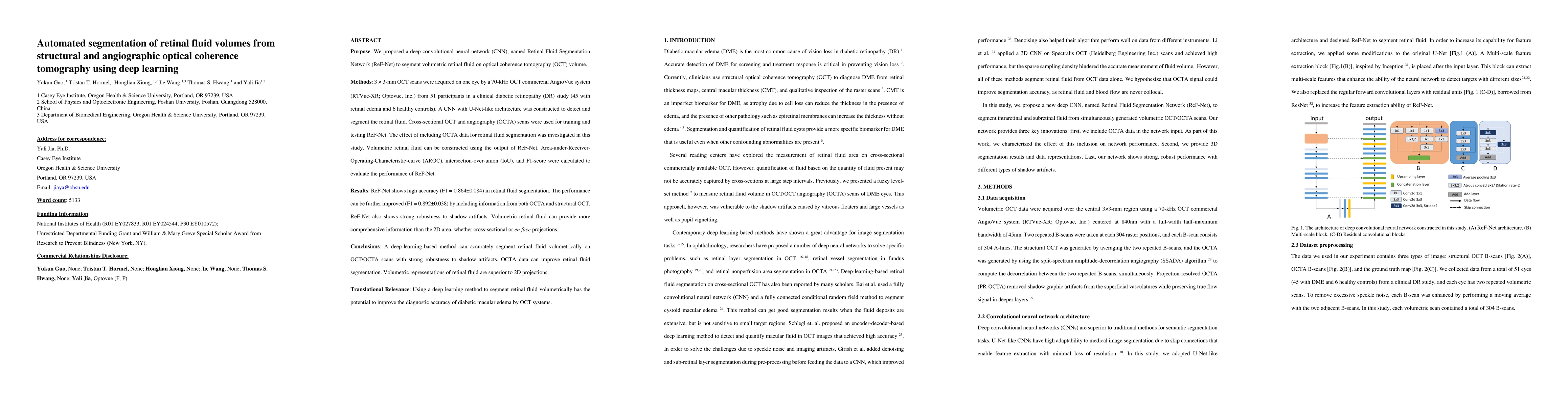
Purpose: We proposed a deep convolutional neural network (CNN), named Retinal Fluid Segmentation Network (ReF-Net) to segment volumetric retinal fluid on optical coherence tomography (OCT) volume. Methods: 3 x 3-mm OCT scans were acquired on one eye by a 70-kHz OCT commercial AngioVue system (RTVue-XR; Optovue, Inc.) from 51 participants in a clinical diabetic retinopathy (DR) study (45 with retinal edema and 6 healthy controls). A CNN with U-Net-like architecture was constructed to detect and segment the retinal fluid. Cross-sectional OCT and angiography (OCTA) scans were used for training and testing ReF-Net. The effect of including OCTA data for retinal fluid segmentation was investigated in this study. Volumetric retinal fluid can be constructed using the output of ReF-Net. Area-under-Receiver-Operating-Characteristic-curve (AROC), intersection-over-union (IoU), and F1-score were calculated to evaluate the performance of ReF-Net. Results: ReF-Net shows high accuracy (F1 = 0.864 +/- 0.084) in retinal fluid segmentation. The performance can be further improved (F1 = 0.892 +/- 0.038) by including information from both OCTA and structural OCT. ReF-Net also shows strong robustness to shadow artifacts. Volumetric retinal fluid can provide more comprehensive information than the 2D area, whether cross-sectional or en face projections. Conclusions: A deep-learning-based method can accurately segment retinal fluid volumetrically on OCT/OCTA scans with strong robustness to shadow artifacts. OCTA data can improve retinal fluid segmentation. Volumetric representations of retinal fluid are superior to 2D projections. Translational Relevance: Using a deep learning method to segment retinal fluid volumetrically has the potential to improve the diagnostic accuracy of diabetic macular edema by OCT systems.

AI Key Findings

Get AI-generated insights about this paper's methodology, results, and significance.

Paper Details

PDF Preview

Download PDF

Key Terms

fluid (0.295)
net (0.279)
segmentation (0.217)
shadow (0.189)
artifacts (0.184)
projections (0.173)
cnn (0.165)
deep (0.141)
robustness (0.140)
deep learning (0.140)
area (0.136)
constructed (0.136)
cross (0.121)
healthy (0.119)
improve (0.118)
en (0.117)
shows (0.112)
accuracy (0.112)
union (0.112)
commercial (0.109)
Title Authors Year Actions
Advances in OCT Angiography
T. Hormel, David Huang, Yali Jia
2025
View Paper
Classifying the severity of diabetic macular oedema from optical coherence tomography scans using deep learning: a feasibility study.
C. L. Breathnach, F. Harney, D. Townley, R. Hickey, A. Simpkin, D. O'Keeffe
2025
View Paper
Denoising, segmentation and volumetric rendering of optical coherence tomography angiography (OCTA) image using deep learning techniques: a review
Kejie Chen, Xiaochun Yang, Jing Na, Wenbo Wang
2025
View Paper
Precision Segmentation of Subretinal Fluids in OCT Using Multiscale Attention-Based U-Net Architecture
P. Karn, Waleed H. Abdulla
2024
View Paper
Novel Method to Measure Volumes of Retinal Specific Entities
Myrta Lippera, Mariantonia Ferrara, Karina Spiess, Nouf Alnafisee, Naseer Ally, A. Jalil, T. Ivanova, George Moussa
2024
View Paper
A robust segmentation of retinal fluids from OCT images using MCFAR-net
Geetha Pavani Pappu, B. Biswal, Srinivasa Rao Kandula, P. Biswal, G. Siddartha, T. Niranjan, N. BalaSubrahmanyam
2024
View Paper
DISCOVER: 2-D Multiview Summarization of Optical Coherence Tomography Angiography for Automatic Diabetic Retinopathy Diagnosis
Mostafa EL HABIB DAHO, Yi-Hsuan Li, Rachid Zeghlache, Hugo Le Boit'e, Pierre Deman, Laurent Borderie, Hugang Ren, Niranchana Mannivanan, Capucine Lepicard, B. Cochener, A. Couturier, R. Tadayoni, Pierre-Henri Conze, M. Lamard, G. Quellec
2024
View Paper
Comparative Analysis of Pre-trained ResNet and DenseNet Models for the Detection of Diabetic Macular Edema
K. Pavithra, Preetham Kumar, M. Geetha, S. Bhandary
2023
View Paper
Alterations of outer retinal reflectivity in diabetic patients without clinically detectable retinopathy.
Feng Zhang, Zijing Du, Xiaying Zhang, Yaxin Wang, Yesheng Chen, Guanrong Wu, Yingying Liang, Dan Cao, Jun Zhao, Ying Fang, Jianhua Ma, Honghua Yu, Yijun Hu
2023
View Paper
Deep-Learning-Based Visualization and Volumetric Analysis of Fluid Regions in Optical Coherence Tomography Scans
Harishwar Reddy Kasireddy, Udaykanth Reddy Kallam, Sowmitri Karthikeya Siddhartha Mantrala, Hemanth Kongara, Anshul Shivhare, Jayesh Saita, Sharanya Vijay, Raghuram Prasad, R. Raman, C. Seelamantula
2023
View Paper
The Present and Future of Artificial Intelligence-Based Medical Image in Diabetes Mellitus: Focus on Analytical Methods and Limitations of Clinical Use
Ji Chun, Hun‐Sung Kim
2023
View Paper
Optical coherence tomography angiography in diabetic retinopathy
N. Waheed, R. Rosen, Yali Jia, M. Munk, David Huang, A. Fawzi, V. Chong, Q. D. Nguyen, Y. Sepah, E. Pearce
2023
View Paper
RGC-Net: An Automatic Reconstruction and Quantification Algorithm for Retinal Ganglion Cells Based on Deep Learning
Rui Ma, Lili Hao, Yudong Tao, Ximena Mendoza, Mohamed M. Khodeiry, Yuan Liu, Mei-Ling Shyu, Richard K. Lee
2023
View Paper
Optical Coherence Tomography Angiography in Retinal Vascular Disorders
Charles Ong, M. Wong, K. X. Cheong, Jinzhi Zhao, K. Teo, Tien-En Tan
2023
View Paper
Automated Macular Fluid Volume As a Treatment Indicator for Diabetic Macular Edema
Kotaro Tsuboi, Q. You, Yukun Guo, Jie Wang, C. Flaxel, Steven T. Bailey, David Huang, Yali Jia, T. Hwang
2023
View Paper
On Machine Learning in Clinical Interpretation of Retinal Diseases Using OCT Images
P. Karn, W. Abdulla
2023
View Paper
Perfused and Nonperfused Microaneurysms Identified and Characterized by Structural and Angiographic OCT
Min Gao, T. Hormel, Yukun Guo, Kotaro Tsuboi, C. Flaxel, David Huang, T. Hwang, Yali Jia
2023
View Paper
A vision transformer architecture for the automated segmentation of retinal lesions in spectral domain optical coherence tomography images
Daniel Philippi, K. Rothaus, Mauro Castelli
2023
View Paper
Deep Learning in Optical Coherence Tomography Angiography: Current Progress, Challenges, and Future Directions
Dawei Yang, A. Ran, Truong X. Nguyen, Timothy P. H. Lin, Haoxing Chen, T. Lai, C. Tham, C. Cheung
2023
View Paper
Computer aided diagnosis of diabetic macular edema in retinal fundus and OCT images: A review
K. Pavithra, Preetham Kumar, M. Geetha, S. Bhandary
2023
View Paper
EANet: Multiscale autoencoder based edge attention network for fluid segmentation from SD‐OCT images
Geetha Pavani Pappu, Sreekar Tankala, Krishna Talabhaktula, B. Biswal
2022
View Paper
Interpretable Diabetic Retinopathy Diagnosis Based on Biomarker Activation Map
P. Zang, T. Hormel, Jie Wang, Yukun Guo, Steven T. Bailey, C. Flaxel, David Huang, T. Hwang, Yali Jia
2022
View Paper
THREE-DIMENSIONAL QUANTIFICATION OF INTRARETINAL CYSTOID SPACES ASSOCIATED WITH FULL-THICKNESS MACULAR HOLE
Kotaro Tsuboi, Yukun Guo, Jie Wang, Elizabeth White, Sam Mershon, M. Kamei, David Huang, Yali Jia, T. Hwang, Steven T. Bailey
2022
View Paper
Evaluation of OCT biomarker changes in treatment-naive neovascular AMD using a deep semantic segmentation algorithm
B. Asani, Olle Holmberg, Johannes Schiefelbein, Michael Hafner, T. Herold, Hannah Spitzer, J. Siedlecki, Christoph Kern, Karsten Kortuem, Amit Frishberg, Fabian J. Theis, S. Priglinger
2022
View Paper
Association Between Visual Acuity and Residual Retinal Fluid Following Intravitreal Anti-Vascular Endothelial Growth Factor Treatment for Neovascular Age-Related Macular Degeneration: A Systematic Review and Meta-analysis.
Nikhil S. Patil, Andrew Mihalache, Arjan S. Dhoot, M. Popovic, R. Muni, P. Kertes
2022
View Paper
Artificial intelligence-based strategies to identify patient populations and advance analysis in age-related macular degeneration clinical trials.
A. Yaghy, Aaron Y. Lee, P. Keane, T. Keenan, L. S. Mendonça, Cecilia S. Lee, A. Cairns, Joseph Caroll, Hao Chen, Julie Clark, C. Cukras, L. de Sisternes, A. Domalpally, M. Durbin, Kerry Goetz, F. Grassmann, J. Haines, Naoto Honda, Z. Hu, Christopher Mody, Luz D. Orozco, C. Owsley, Stephen H Poor, Charles A. Reisman, Ramiro Ribeiro, S. Sadda, S. Sivaprasad, G. Staurenghi, D. Ting, S. Tumminia, Luca Zalunardo, N. Waheed
2022
View Paper
Three-dimensional diabetic macular edema thickness maps based on fluid segmentation and fovea detection using deep learning.
Jingjing Xu, Yang Zhou, Qianyue Wei, Kang Li, Zhenping Li, T. Yu, Jianchun Zhao, Dayong Ding, Xirong Li, Guangze Wang, H. Dai
2022
View Paper
Directional analysis of intensity changes for determining the existence of cyst in optical coherence tomography images
M. Monemian, H. Rabbani
2022
View Paper
ADC-Net: An Open-Source Deep Learning Network for Automated Dispersion Compensation in Optical Coherence Tomography
Shahjalal Ahmed, David Le, Taeyoon Son, Tobiloba Adejumo, Xincheng Yao
2022
View Paper
An Open-Source Deep Learning Network for Reconstruction of High-Resolution OCT Angiograms of Retinal Intermediate and Deep Capillary Plexuses
Min Gao, T. Hormel, Jie Wang, Yukun Guo, Steven T. Bailey, T. Hwang, Yali Jia
2021
View Paper
Open-source deep learning-based automatic segmentation of mouse Schlemm's canal in optical coherence tomography images.
K. C. Choy, Guorong Li, W. Stamer, Sina Farsiu
2021
View Paper
Towards data-driven filters in Paraview
Drishti Maharjan, Peter Zaspel
2021
View Paper
Machine learning in optical coherence tomography angiography
David Le, Taeyoon Son, Xincheng Yao
2021
View Paper
An end-to-end network for segmenting the vasculature of three retinal capillary plexuses from OCT angiographic volumes.
Yukun Guo, T. Hormel, Shaohua Pi, Xiang Wei, Min Gao, J. Morrison, Yali Jia
2021
View Paper
AI-based monitoring of retinal fluid in disease activity and under therapy
U. Schmidt-Erfurth, G. Reiter, Sophie Riedl, Philipp Seeböck, Wolf-Dieter Vogl, B. Blodi, A. Domalpally, A. Fawzi, Yali Jia, D. Sarraf, H. Bogunović
2021
View Paper
Artificial intelligence for improving sickle cell retinopathy diagnosis and management
Sophie Cai, Ian C. Han, A. Scott
2021
View Paper
Automated Quantification of Pathological Fluids in Neovascular Age-Related Macular Degeneration, and Its Repeatability Using Deep Learning
I. Mantel, Agata Mosinska, C. Bergin, M. S. Polito, J. Guidotti, S. Apostolopoulos, Carlos Ciller, Sandro De Zanet
2021
View Paper
DEFN: Dual-Encoder Fourier Group Harmonics Network for Three-Dimensional Macular Hole Reconstruction with Stochastic Retinal Defect Augmentation and Dynamic Weight Composition
Xingru Huang, Yihao Guo, Jian Huang, Zhi Li, Tianyun Zhang, Kunyan Cai, Gaopeng Huang, Wenhao Chen, Zhaoyang Xu, Liangqiong Qu, Ji Hu, Tinyu Wang, Shaowei Jiang, C. Yan, Yaoqi Sun, Xin Ye, Yaqi Wang
2023
View Paper

Comments (0)

This website uses cookies to ensure you get the best experience. Learn more