Automatic airway segmentation from Computed Tomography using robust and efficient 3-D convolutional neural networks
Publication
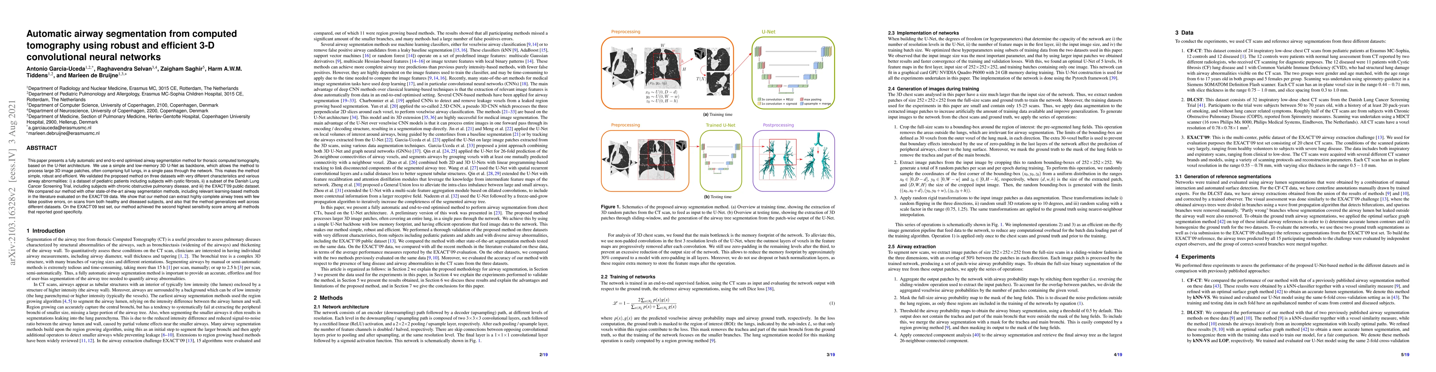
Metrics
AI Quick Summary
This paper introduces a fully automatic airway segmentation method using a 3D U-Net architecture, demonstrating robust and efficient processing of large 3D image patches. The method generalizes well across different datasets and outperforms other state-of-the-art methods in terms of sensitivity and completeness of airway tree extraction.
Paper Preview
Abstract
This paper presents a fully automatic and end-to-end optimised airway segmentation method for thoracic computed tomography, based on the U-Net architecture. We use a simple and low-memory 3D U-Net as backbone, which allows the method to process large 3D image patches, often comprising full lungs, in a single pass through the network. This makes the method simple, robust and efficient. We validated the proposed method on three datasets with very different characteristics and various airway abnormalities: i) a dataset of pediatric patients including subjects with cystic fibrosis, ii) a subset of the Danish Lung Cancer Screening Trial, including subjects with chronic obstructive pulmonary disease, and iii) the EXACT'09 public dataset. We compared our method with other state-of-the-art airway segmentation methods, including relevant learning-based methods in the literature evaluated on the EXACT'09 data. We show that our method can extract highly complete airway trees with few false positive errors, on scans from both healthy and diseased subjects, and also that the method generalizes well across different datasets. On the EXACT'09 test set, our method achieved the second highest sensitivity score among all methods that reported good specificity.
AI Key Findings
Get AI-generated insights about this paper's methodology, results, significance, and more — seven facets brought into focus.
Impact
Paper Details
PDF Preview
Key Terms
Citation Network
Current paper (gray), citations (green), references (blue)
Display is limited for performance on very large graphs.
Discussion 0