Summary
The hippocampus is a seminal structure in the most common surgically-treated form of epilepsy. Accurate segmentation of the hippocampus aids in establishing asymmetry regarding size and signal characteristics in order to disclose the likely site of epileptogenicity. With sufficient refinement, it may ultimately aid in the avoidance of invasive monitoring with its expense and risk for the patient. To this end, a reliable and consistent method for segmentation of the hippocampus from magnetic resonance imaging (MRI) is needed. In this work, we present a systematic and statistical analysis approach for evaluation of automated segmentation methods in order to establish one that reliably approximates the results achieved by manual tracing of the hippocampus.
AI Key Findings
Generated Sep 03, 2025
Methodology
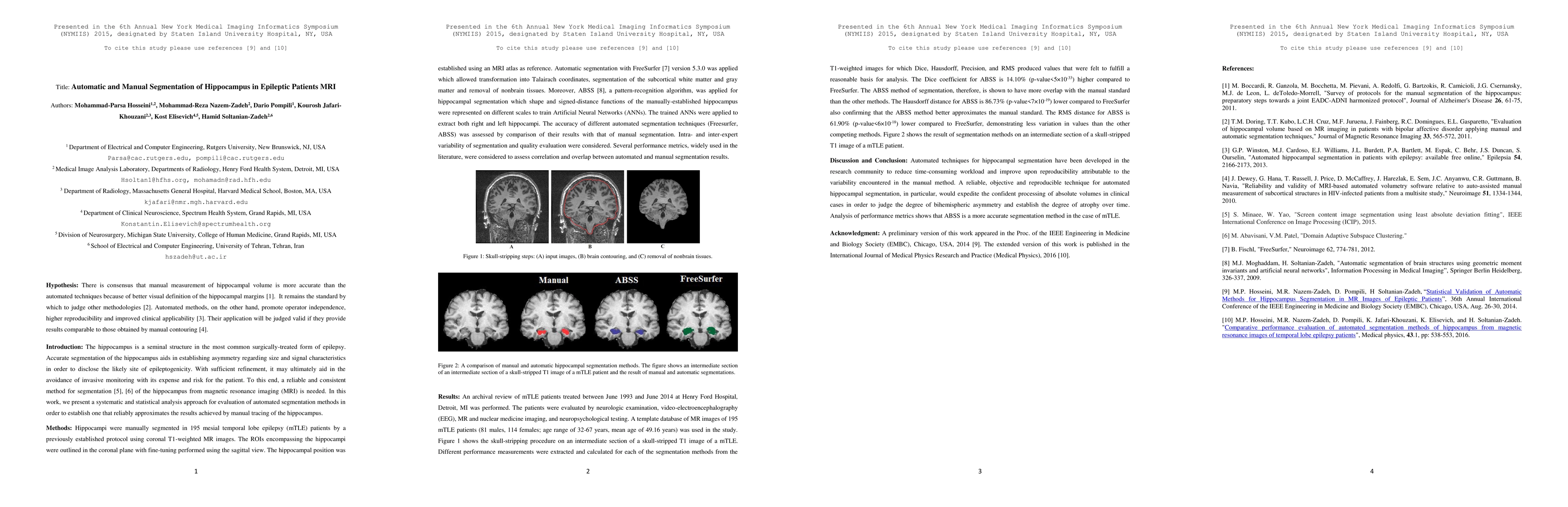
The study manually segmented hippocampi in 195 mesial temporal lobe epilepsy patients using coronal T1-weighted MR images, employing an MRI atlas as a reference. Automated segmentation was performed using FreeSurfer version 5.3.0 and an Artificial Neural Network (ANN)-based pattern-recognition algorithm called ABSS.
Key Results
- ABSS method showed higher Dice coefficient (14.10%) and lower Hausdorff (86.73%) and RMS distances (61.90%) compared to FreeSurfer, indicating better overlap with manual segmentation results.
- Performance metrics (Dice, Hausdorff, Precision, and RMS) demonstrated that ABSS is a more accurate segmentation method for mTLE patients.
- The study concluded that ABSS outperforms FreeSurfer in automated hippocampal segmentation for mTLE patients.
Significance
This research is important as it aims to develop a reliable, objective, and reproducible technique for automated hippocampal segmentation, which can expedite clinical processing and judge the degree of bihemispheric asymmetry and atrophy over time, ultimately aiding in avoiding invasive monitoring for epilepsy patients.
Technical Contribution
The main technical contribution of this work is the development and validation of an ANN-based pattern-recognition algorithm (ABSS) for hippocampal segmentation, which outperforms the widely-used FreeSurfer method in terms of accuracy and overlap with manual segmentation results.
Novelty
This research presents a novel approach using ABSS, an ANN-based method, for automated hippocampal segmentation, which demonstrates superior performance compared to the established FreeSurfer technique, addressing the need for a reliable and consistent method for hippocampal segmentation from MRI in epilepsy patients.
Limitations
- The study was limited to mTLE patients and did not explore the applicability of the methods to other epilepsy types or neurological conditions.
- The manual segmentation process might still introduce inter- and intra-expert variability, which could affect the overall accuracy of automated methods.
Future Work
- Expand the study to other epilepsy types and neurological conditions to assess the generalizability of the ABSS method.
- Investigate methods to reduce inter- and intra-expert variability in manual segmentation to improve automated method validation.
Paper Details
PDF Preview
Key Terms
Citation Network
Current paper (gray), citations (green), references (blue)
Display is limited for performance on very large graphs.
Similar Papers
Found 4 papersDilated deeply supervised networks for hippocampus segmentation in MRI
Andreas Maier, Nishant Ravikumar, Sulaiman Vesal et al.
| Title | Authors | Year | Actions |
|---|
Comments (0)