Automatic Aortic Valve Pathology Detection from 3-Chamber Cine MRI with Spatio-Temporal Attention Maps
Publication
Metrics
AI Quick Summary
This paper proposes a 3D neural network to automatically classify aortic valve pathology from 3-chamber cine MRI scans, achieving an accuracy of 0.85 +/- 0.03 for detection and 0.75 +/- 0.03 for discrimination of specific types. The model highlights the importance of blood pool voxels near the aortic root for accurate diagnosis.
Paper Preview
Abstract
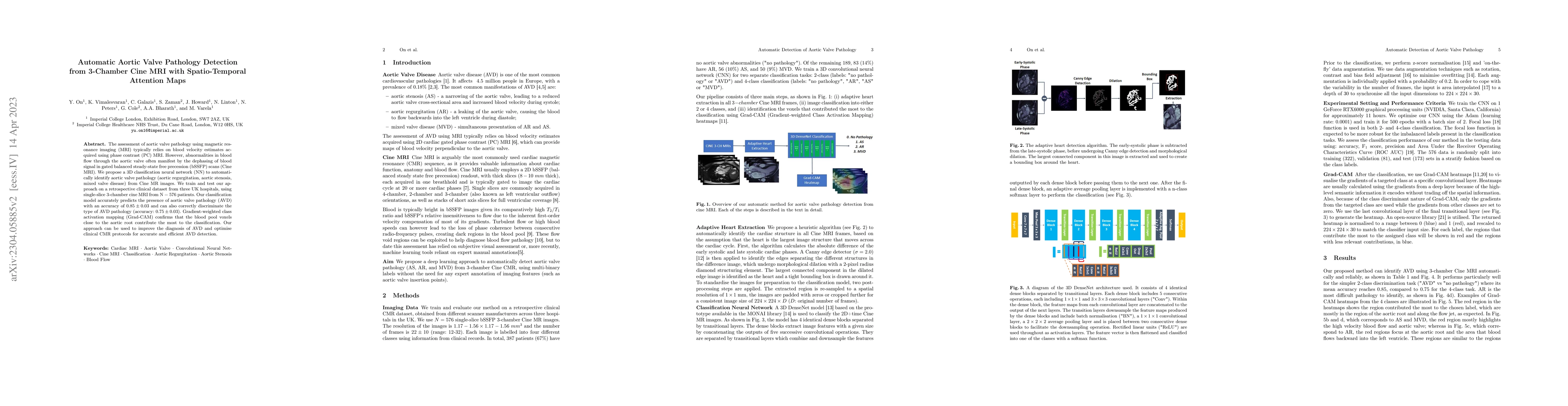
The assessment of aortic valve pathology using magnetic resonance imaging (MRI) typically relies on blood velocity estimates acquired using phase contrast (PC) MRI. However, abnormalities in blood flow through the aortic valve often manifest by the dephasing of blood signal in gated balanced steady-state free precession (bSSFP) scans (Cine MRI). We propose a 3D classification neural network (NN) to automatically identify aortic valve pathology (aortic regurgitation, aortic stenosis, mixed valve disease) from Cine MR images. We train and test our approach on a retrospective clinical dataset from three UK hospitals, using single-slice 3-chamber cine MRI from N = 576 patients. Our classification model accurately predicts the presence of aortic valve pathology (AVD) with an accuracy of 0.85 +/- 0.03 and can also correctly discriminate the type of AVD pathology (accuracy: 0.75 +/- 0.03). Gradient-weighted class activation mapping (Grad-CAM) confirms that the blood pool voxels close to the aortic root contribute the most to the classification. Our approach can be used to improve the diagnosis of AVD and optimise clinical CMR protocols for accurate and efficient AVD detection.
AI Key Findings
Get AI-generated insights about this paper's methodology, results, significance, and more — seven facets brought into focus.
Impact
Paper Details
Authors
PDF Preview
Key Terms
Citation Network
Current paper (gray), citations (green), references (blue)
Display is limited for performance on very large graphs.
Discussion 0