Summary
Rapid and reliable vascular access is critical in trauma and critical care. Central vascular catheterization enables high-volume resuscitation, hemodynamic monitoring, and advanced interventions like ECMO and REBOA. While peripheral access is common, central access is often necessary but requires specialized ultrasound-guided skills, posing challenges in prehospital settings. The complexity arises from deep target vessels and the precision needed for needle placement. Traditional techniques, like the Seldinger method, demand expertise to avoid complications. Despite its importance, ultrasound-guided central access is underutilized due to limited field expertise. While autonomous needle insertion has been explored for peripheral vessels, only semi-autonomous methods exist for femoral access. This work advances toward full automation, integrating robotic ultrasound for minimally invasive emergency procedures. Our key contribution is the successful femoral vein and artery cannulation in a porcine hemorrhagic shock model.
AI Key Findings
Generated Sep 02, 2025
Methodology
The research utilized a robotic system consisting of a Universal Robots UR3 arm, a Fukuda linear ultrasound probe, a 6-axis ATI Nano 25 force sensor, an Intel RealSense camera, and a needle insertion mechanism. The system was tested on live porcine subjects under anesthesia, with a midline laparotomy followed by stellate through-and-through liver lacerations to model non-compressible hemorrhage. Robotic cannulation of femoral vein and artery was performed once the mean arterial pressure (MAP) dropped below 40 mmHg.
Key Results
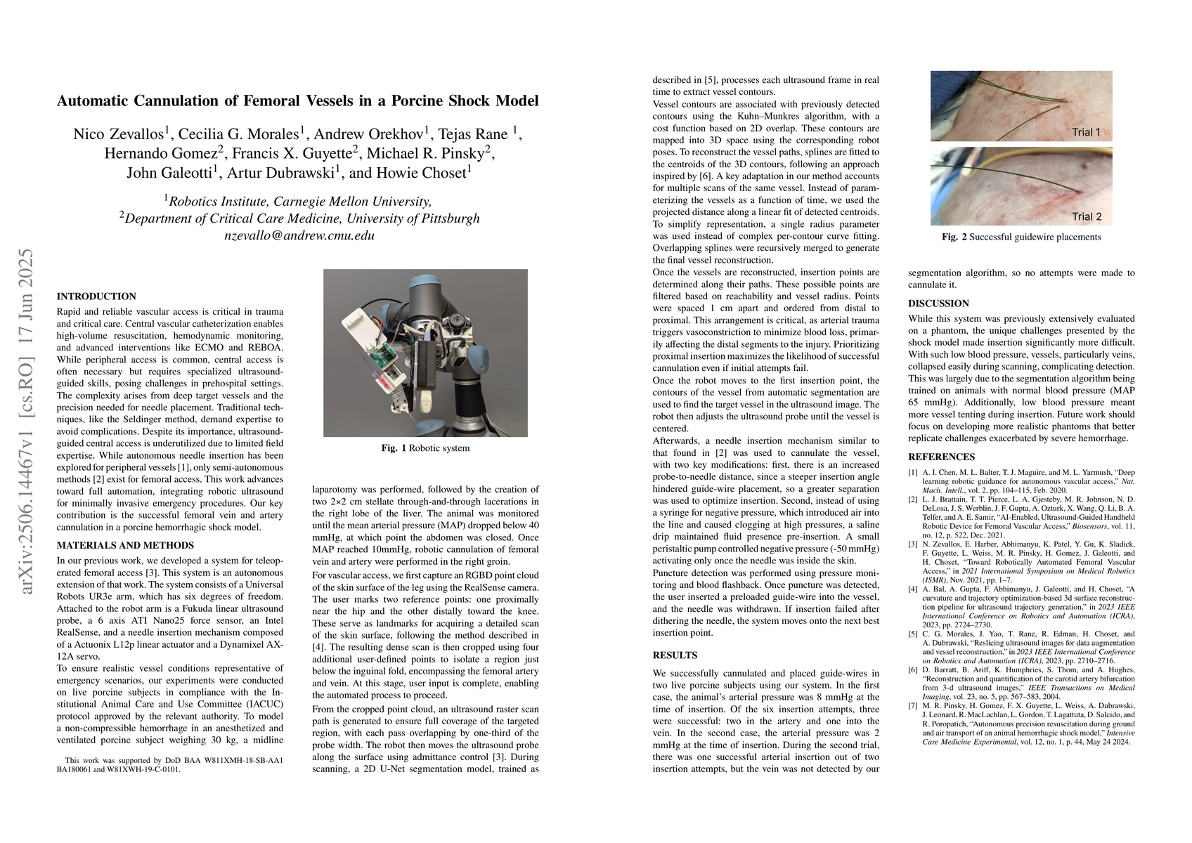
- Successful femoral vein and artery cannulation in a porcine hemorrhagic shock model.
- Three successful insertions out of six attempts in the first porcine subject (two in artery, one in vein).
- One successful arterial insertion out of two attempts in the second porcine subject.
Significance
This research advances toward full automation of minimally invasive emergency procedures, addressing the challenges of ultrasound-guided central access in prehospital settings due to limited field expertise.
Technical Contribution
Integration of robotic ultrasound for automated femoral vessel cannulation in a porcine shock model, including automated vessel segmentation, insertion point determination, and guide-wire placement.
Novelty
This work distinguishes itself by focusing on full automation for femoral vessel cannulation, unlike existing semi-autonomous methods, and by addressing the unique challenges presented by a porcine hemorrhagic shock model.
Limitations
- The segmentationalgorithm was trained on animals with normal blood pressure, making it less effective in detecting collapsed vessels in low-pressure scenarios.
- Low blood pressure led to more vessel tenting during insertion, complicating the cannulation process.
Future Work
- Develop more realistic phantom models to better replicate challenges exacerbated by severe hemorrhage.
- Improve the algorithm's performance in low-pressure conditions.
Paper Details
PDF Preview
Citation Network
Current paper (gray), citations (green), references (blue)
Display is limited for performance on very large graphs.
Similar Papers
Found 4 papersA Feasible Workflow for Retinal Vein Cannulation in Ex Vivo Porcine Eyes with Robotic Assistance
Marin Kobilarov, Iulian Iordachita, Peter Gehlbach et al.
No citations found for this paper.
Comments (0)