Automatic Classification of Bright Retinal Lesions via Deep Network Features
Publication
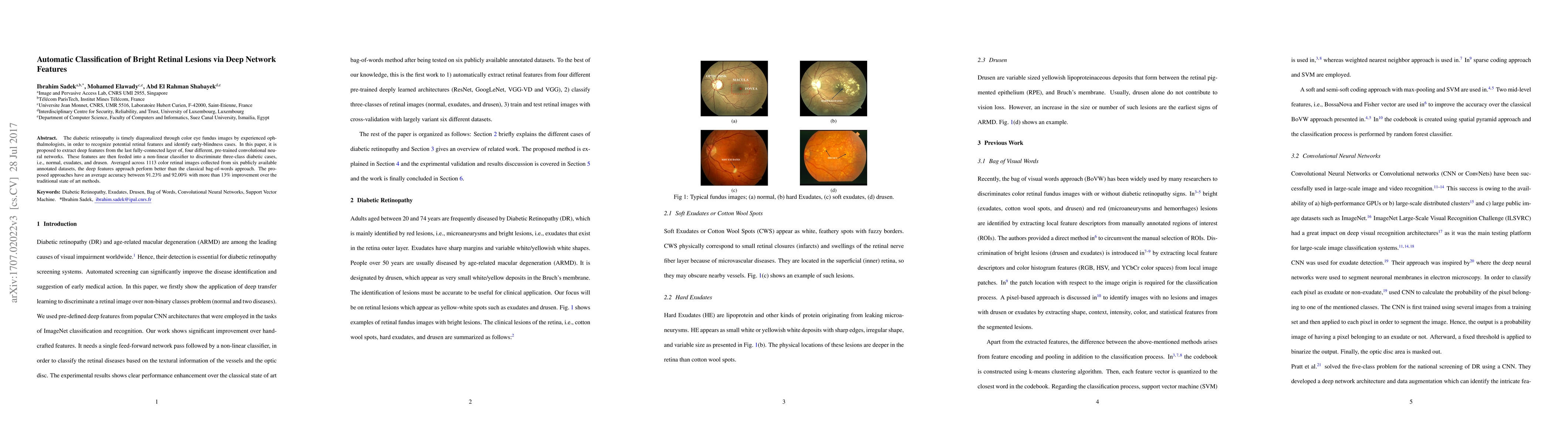
Metrics
AI Quick Summary
This paper proposes using deep features from pre-trained convolutional neural networks to classify diabetic retinopathy into normal, exudates, and drusen categories. The method outperforms traditional bag-of-words approaches, achieving an average accuracy of 91.23% to 92.00% across 1113 retinal images.
Paper Preview
Abstract
The diabetic retinopathy is timely diagonalized through color eye fundus images by experienced ophthalmologists, in order to recognize potential retinal features and identify early-blindness cases. In this paper, it is proposed to extract deep features from the last fully-connected layer of, four different, pre-trained convolutional neural networks. These features are then feeded into a non-linear classifier to discriminate three-class diabetic cases, i.e., normal, exudates, and drusen. Averaged across 1113 color retinal images collected from six publicly available annotated datasets, the deep features approach perform better than the classical bag-of-words approach. The proposed approaches have an average accuracy between 91.23% and 92.00% with more than 13% improvement over the traditional state of art methods.
AI Key Findings
Get AI-generated insights about this paper's methodology, results, significance, and more — seven facets brought into focus.
Impact
Paper Details
PDF Preview
Key Terms
Citation Network
Current paper (gray), citations (green), references (blue)
Display is limited for performance on very large graphs.
Discussion 0