This paper proposes a wavelet-based statistical texture analysis method for automatic diagnosis of abnormal tumor regions in brain CT images. The system uses Discrete Wavelet Decomposition, feature extraction, selection via Genetic Algorithm, and classification with SVM, achieving 96% accuracy, outperforming BPN classifiers.
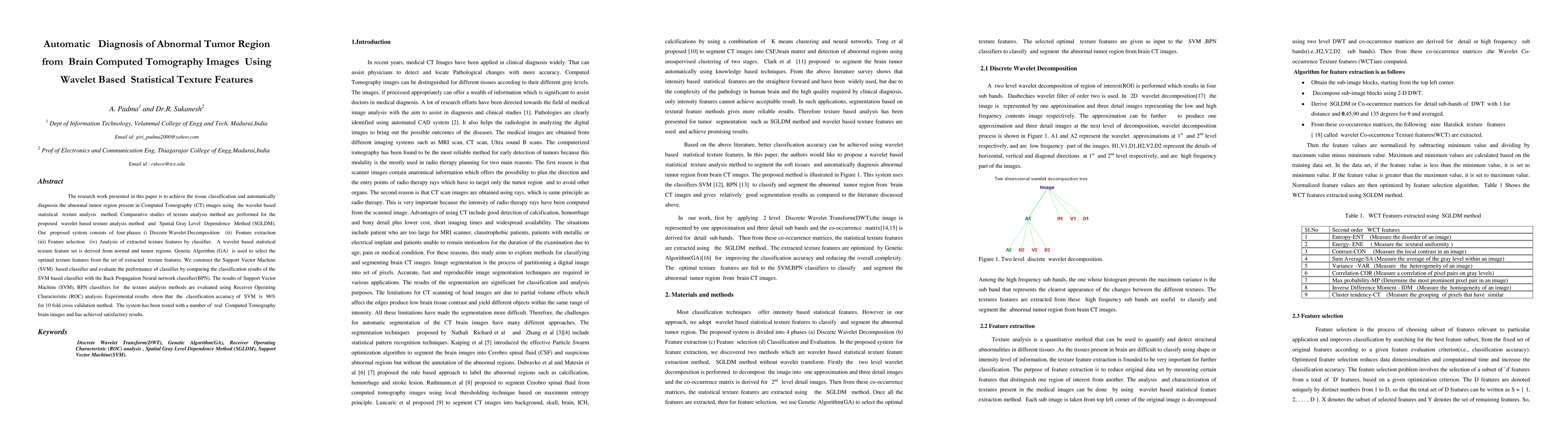
The research work presented in this paper is to achieve the tissue classification and automatically diagnosis the abnormal tumor region present in Computed Tomography (CT) images using the wavelet based statistical texture analysis method. Comparative studies of texture analysis method are performed for the proposed wavelet based texture analysis method and Spatial Gray Level Dependence Method (SGLDM). Our proposed system consists of four phases i) Discrete Wavelet Decomposition (ii) Feature extraction (iii) Feature selection (iv) Analysis of extracted texture features by classifier. A wavelet based statistical texture feature set is derived from normal and tumor regions. Genetic Algorithm (GA) is used to select the optimal texture features from the set of extracted texture features. We construct the Support Vector Machine (SVM) based classifier and evaluate the performance of classifier by comparing the classification results of the SVM based classifier with the Back Propagation Neural network classifier(BPN). The results of Support Vector Machine (SVM), BPN classifiers for the texture analysis methods are evaluated using Receiver Operating Characteristic (ROC) analysis. Experimental results show that the classification accuracy of SVM is 96% for 10 fold cross validation method. The system has been tested with a number of real Computed Tomography brain images and has achieved satisfactory results.
This graph shows how this paper is connected to other research. The current paper is shown in dark gray, papers that cite it are in green, and papers it references are in blue. You can:
• Hover over any node to see the paper title
• Click a node to view that paper's details
• Drag nodes to rearrange the graph
• Use the zoom controls (bottom right) to zoom in/out
Title | Authors | Year | Actions |
---|---|---|---|
Fractional probabilistic fuzzy clustering and optimization based brain tumor segmentation and classification
|
T. A. Jemimma, Y. J. V. Raj
|
2022
|
View Paper |
Particle Rider Mutual Information and Dendritic-Squirrel Search Algorithm With Artificial Immune Classifier for Brain Tumor Classification
|
R. R. Chakre, D. Patil
|
2021
|
View Paper |
Imaging Analysis of Thresholding Image Filtering, Brain Abnormalities Morphology, and Dose Report CT Scan Records
|
Bunga Meyzia, M. Hamdi, Saktioto, R. Amelia
|
2020
|
View Paper |
Classification of Brain Tumors Using Convolutional Neural Network over Various SVM Methods
|
V. R. Sajja, H. Kalluri
|
2020
|
View Paper |
Decision fusion scheme for bearing defects diagnosis in induction motors
|
H. Agahi, A. Mahmoodzadeh
|
2020
|
View Paper |
Diagnosis and Classification of Brain Tumor
|
Ravendra Singh
|
2019
|
View Paper |
A novel intensity-based multi-level classification approach for coronary plaque characterization in intravascular ultrasound images
|
G. Kim, Ju Hwan Lee, Y. Hwang, Sung Min Kim
|
2018
|
View Paper |
An Effective Brain Tumor Diagnosis and Monitoring By Spreading Area Measurement
|
Jisna Jose, D. Anto, Sahaya Dhas
|
2017
|
View Paper |
DWT based Feature Extraction for Classification of Untreated MRI Mammogram of Breast Cells and Normal Cells
|
S. Sushma, S. Balasubramanian, K. Latha
|
2017
|
View Paper |
Histogram-Based Texture Characterization and Classification of Brain Tissues in Non-Contrast CT Images of Stroke Patients
|
K. Agwu, C. Ohagwu
|
2016
|
View Paper |
Brain tumour classification using two-tier classifier with adaptive segmentation technique
|
V. Anitha, S. Murugavalli
|
2016
|
View Paper |
Performance evaluation of support vector machine and artificial neural network in the classification of liver cirhosis and hemachromatosis
|
O. Fenwa, F. Ajala, A. Aku
|
2015
|
View Paper |
Classification of medical images using the model for formation of viewing trajectory
|
V. Osinov, E. S. Pel’meneva, D. Shaposhnikov
|
2015
|
View Paper |
Leather texture classification using wavelet feature extraction technique
|
M. Jawahar, N. K. Chandra Babu, K. Vani
|
2014
|
View Paper |
PRE-SEGMENTATION F OR THE COMPUTER AIDED DIAGNOSIS SYSTEM
|
M. Kyaw, Pyin Oo Lwin
|
2013
|
View Paper |
Brain Tumor detection and identification in brain MRI using supervised learning: A LDA based classification method
|
2017
|
View Paper | |
DWT based Feature Extraction for Classification of Untreated MRI Mammogram of Breast Cells and Normal Cells
|
E. Rashed, I. A. Ismail, S. Zaki
|
2017
|
View Paper |
A Study on Optimization Techniques in Feature Selection for Medical Image Analysis
|
M. Allam, Dr. M. Nandhini
|
2017
|
View Paper |
Brain Tumor Classification Using Machine Learning Algorithms
|
B.Balakumar, P.Raviraj, E.Divya
|
2017
|
View Paper |
Prostate Disease Diagnosis from CT Images Using GA Optimized SMRT Based Texture Features
|
B. Manju, K. Meenakshy, R. Gopikakumari
|
2015
|
View Paper |
FUZZY BASED AUTOMATIC DETECTION AND CLASSIFICATION APPROACH FOR MRI-BRAIN TUMOR
|
R. Karuppathal, V. Palanisamy
|
2014
|
View Paper |
Evaluating the Hausdorff Distance for Contour Segmentation of Brain Images
|
N. Kamal, T. Nadu
|
2014
|
View Paper |
Computer-Aided Detection system for Hemorrhage contained region
|
M. Kyaw, Yatanarpon Cybercity, Pyin Oo Lwin
|
2013
|
View Paper |
An Efficient MR Image Brain Tumor Segmentation Based on Discrete Wavelet Transform and Region Growing Algorithm
|
V. Ramya
|
2013
|
View Paper |
Automatic Medical Image Classification and Abnormality Detection Using K-Nearest Neighbour
|
Dr. R. J. Ramteke, Khachane Monali
|
2012
|
View Paper |
Published: 2019-06-25
Published: 2015-12-29
Vidhyapriya Ranganathan, Celshiya Udaiyar, Jaisree Jayanth, Meghaa P V, Srija B, Uthra S
Published: 2024-07-15
Comments (0)