Summary
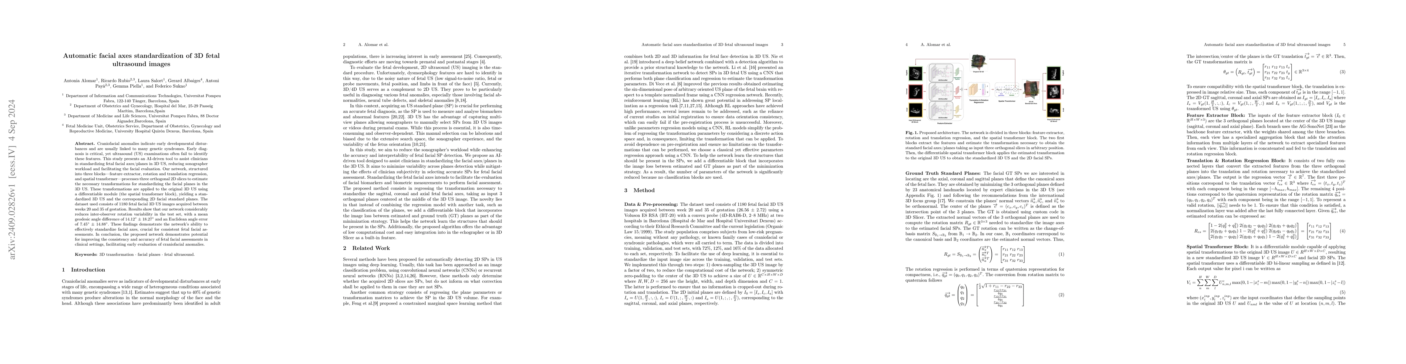
Craniofacial anomalies indicate early developmental disturbances and are usually linked to many genetic syndromes. Early diagnosis is critical, yet ultrasound (US) examinations often fail to identify these features. This study presents an AI-driven tool to assist clinicians in standardizing fetal facial axes/planes in 3D US, reducing sonographer workload and facilitating the facial evaluation. Our network, structured into three blocks-feature extractor, rotation and translation regression, and spatial transformer-processes three orthogonal 2D slices to estimate the necessary transformations for standardizing the facial planes in the 3D US. These transformations are applied to the original 3D US using a differentiable module (the spatial transformer block), yielding a standardized 3D US and the corresponding 2D facial standard planes. The dataset used consists of 1180 fetal facial 3D US images acquired between weeks 20 and 35 of gestation. Results show that our network considerably reduces inter-observer rotation variability in the test set, with a mean geodesic angle difference of 14.12$^{\circ}$ $\pm$ 18.27$^{\circ}$ and an Euclidean angle error of 7.45$^{\circ}$ $\pm$ 14.88$^{\circ}$. These findings demonstrate the network's ability to effectively standardize facial axes, crucial for consistent fetal facial assessments. In conclusion, the proposed network demonstrates potential for improving the consistency and accuracy of fetal facial assessments in clinical settings, facilitating early evaluation of craniofacial anomalies.
AI Key Findings
Get AI-generated insights about this paper's methodology, results, and significance.
Paper Details
PDF Preview
Citation Network
Current paper (gray), citations (green), references (blue)
Display is limited for performance on very large graphs.
No citations found for this paper.
Comments (0)