Automatic Grading of Individual Knee Osteoarthritis Features in Plain Radiographs using Deep Convolutional Neural Networks

Source: ArXiv

AI Quick Summary

This study developed a deep convolutional neural network to automatically grade knee osteoarthritis features in radiographs, achieving high accuracy in predicting both Kellgren-Lawrence and OARSI grades, outperforming current state-of-the-art methods.

Share: Share on X LinkedIn

Summary

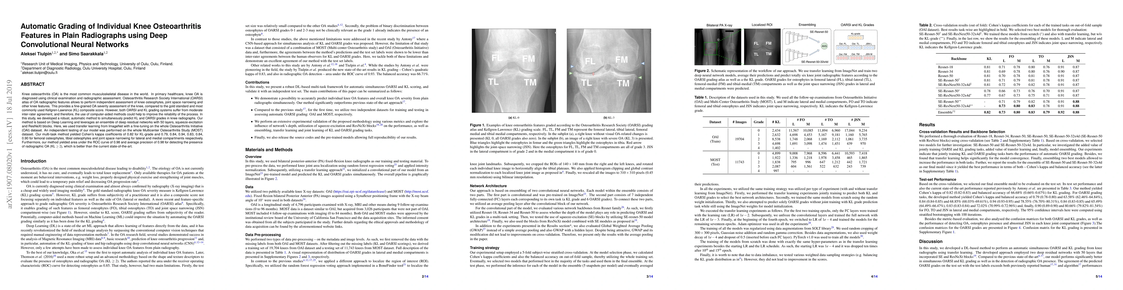
Knee osteoarthritis (OA) is the most common musculoskeletal disease in the world. In primary healthcare, knee OA is diagnosed using clinical examination and radiographic assessment. Osteoarthritis Research Society International (OARSI) atlas of OA radiographic features allows to perform independent assessment of knee osteophytes, joint space narrowing and other knee features. This provides a fine-grained OA severity assessment of the knee, compared to the gold standard and most commonly used Kellgren-Lawrence (KL) composite score. However, both OARSI and KL grading systems suffer from moderate inter-rater agreement, and therefore, the use of computer-aided methods could help to improve the reliability of the process. In this study, we developed a robust, automatic method to simultaneously predict KL and OARSI grades in knee radiographs. Our method is based on Deep Learning and leverages an ensemble of deep residual networks with 50 layers, squeeze-excitation and ResNeXt blocks. Here, we used transfer learning from ImageNet with a fine-tuning on the whole Osteoarthritis Initiative (OAI) dataset. An independent testing of our model was performed on the whole Multicenter Osteoarthritis Study (MOST) dataset. Our multi-task method yielded Cohen's kappa coefficients of 0.82 for KL-grade and 0.79, 0.84, 0.94, 0.83, 0.84, 0.90 for femoral osteophytes, tibial osteophytes and joint space narrowing for lateral and medial compartments respectively. Furthermore, our method yielded area under the ROC curve of 0.98 and average precision of 0.98 for detecting the presence of radiographic OA (KL $\geq 2$), which is better than the current state-of-the-art.

AI Key Findings

Generated Sep 02, 2025

Methodology

The research developed a deep learning-based method using transfer learning from ImageNet to automatically grade Kellgren-Lawrence (KL) and Osteoarthritis Research Society International (OARSI) features from knee radiographs, employing deep residual networks with 50 layers incorporating Squeeze-Excitation and ResNeXt blocks.

Key Results

  • The method yielded Cohen's kappa coefficients of 0.82 for KL-grade and 0.79, 0.84, 0.94, 0.83, 0.84, 0.90 for femoral osteophytes, tibial osteophytes, and joint space narrowing for lateral and medial compartments, respectively.
  • Area under the ROC curve of 0.98 and average precision of 0.98 for detecting the presence of radiographic OA (KL ≥ 2) was achieved, surpassing the current state-of-the-art.
  • Cross-validation and testing on independent datasets (OAI and MOST) demonstrated robust performance.

Significance

This research provides a significant advancement in computer-aided diagnosis for knee osteoarthritis, offering improved reliability and fine-grained severity assessment compared to traditional methods, which could benefit clinical trials and routine practice.

Technical Contribution

The development of an ensemble deep learning model combining residual networks with Squeeze-Excitation and ResNeXt blocks, leveraging transfer learning for automatic grading of KL and OARSI features in knee radiographs.

Novelty

This work presents the first large-scale experiment for automatic KL and OARSI grading from plain radiographs, significantly outperforming previous state-of-the-art methods in both detection of radiographic OA presence and individual feature grading.

Limitations

  • The study did not analyze attention maps produced by the model, which could offer further insights into specific decisions made by the CNN.
  • The computational heaviness of the ensemble approach might affect its real-life applicability unless deployed on GPU.
  • The study utilized whole knee images for training and did not compare this approach with the Siamese model proposed by Tiulpin et al.
  • Some OARSI features, like medial tibial attrition, medial tibial sclerosis, and lateral femoral sclerosis, were not considered due to insufficient training and test data.
  • The test set included data from the same patients obtained from multiple follow-ups, which, although not significantly affecting results, made them less optimistic.

Future Work

  • Investigate attention maps to gain deeper understanding of model decisions.
  • Explore techniques like knowledge distillation to reduce computational effort.
  • Compare the whole image approach with the Siamese model for potential improvements.
  • Consider additional OARSI features not currently analyzed.
  • Evaluate the model's performance using data from only the first imaging follow-up in the MOST dataset to validate the impact of using multi-followup data.

Paper Details

PDF Preview

Download PDF

Key Terms

assessment (0.240)
joint (0.176)
fine (0.160)
dataset (0.143)
independent (0.141)
deep (0.136)
method (0.134)
atlas (0.133)
Title Authors Year Actions

Comments (0)

This website uses cookies to ensure you get the best experience. Learn more