ArxivLens

Quick Summary:

This paper proposes a new Local Binary Pattern-based method for automatic identification of retinal arteries and veins in fundus images, emphasizing its robustness against low-contrast and low-quality images. Experimental results show superior performance compared to state-of-the-art and other feature extraction methods.

Journal Reference: Investigative Ophthalmology and Visual Science, 55 (5), 232, 2014
arXiv Id: 1605.00763
Date Published: 2016-05-04
Date Updated: 2016-05-04

Categories:

Key Terms (TF-IDF):

classification (0.272)
classification (0.257)
images (0.209)
images (0.205)
state art (0.192)
reviews (0.192)
art (0.191)
methods (0.191)
state art (0.188)
art (0.187)
feature extraction (0.187)
based method (0.185)
methods (0.185)
compare performance (0.185)
reviews (0.183)
based method (0.183)
texture (0.182)
compare performance (0.181)
helps (0.179)
License: http://arxiv.org/licenses/nonexclusive-distrib/1.0/

Summary:

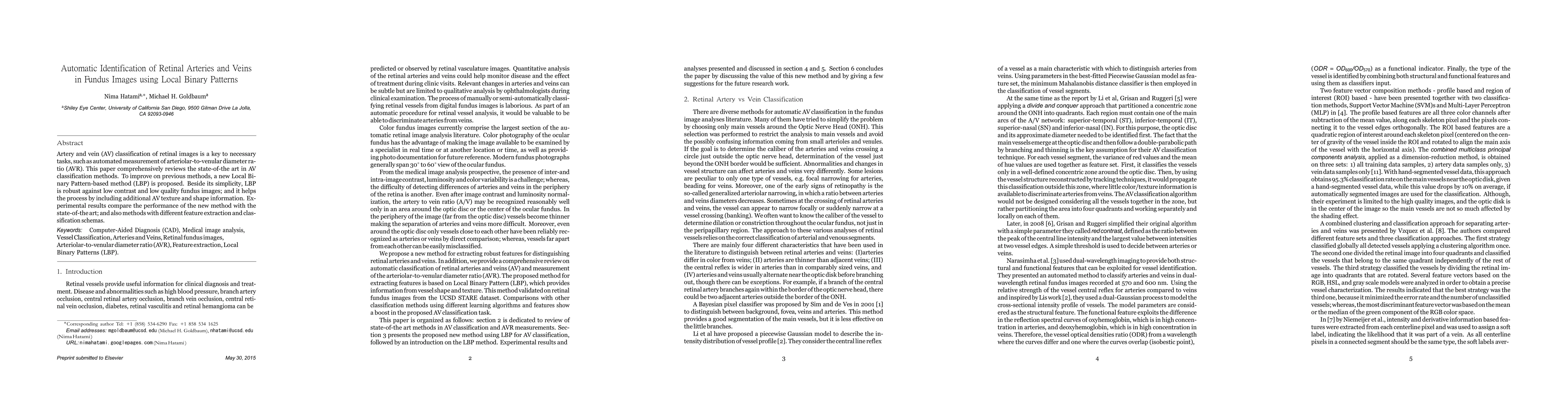
Artery and vein (AV) classification of retinal images is a key to necessary tasks, such as automated measurement of arteriolar-to-venular diameter ratio (AVR). This paper comprehensively reviews the state-of-the art in AV classification methods. To improve on previous methods, a new Local Bi- nary Pattern-based method (LBP) is proposed. Beside its simplicity, LBP is robust against low contrast and low quality fundus images; and it helps the process by including additional AV texture and shape information. Experimental results compare the performance of the new method with the state-of-the art; and also methods with different feature extraction and classification schemas.

Citation & Reference Network Visualization

This graph shows how this paper is connected to other research. The current paper is shown in dark gray, papers that cite it are in green, and papers it references are in blue. You can:

Current Paper
Papers citing this work (Citations)
Papers cited by this work (References)

• Hover over any node to see the paper title

• Click a node to view that paper's details

• Drag nodes to rearrange the graph

• Use the zoom controls (bottom right) to zoom in/out

Citations and References

Title Authors Year Actions
Automated analysis of vessel morphometry in retinal images from a Danish high street optician setting
J. Freiberg, R. Welikala, Jens Rovelt, C. Owen, A. Rudnicka, M. Kolko, S. Barman
2023
View Paper
Diabetic Retinopathy Diagnosis Through Computer-Aided Fundus Image Analysis: A Review
Jaskirat Kaur, D. Mittal, Ruchika Singla
2021
View Paper
Retinal Blood Vessel Segmentation Using Pix2Pix GAN*
D. Popescu, Mihaela Deaconu, L. Ichim, G. Stamatescu
2021
View Paper
Diameter Estimation of Fallopian Tubes Using Visual Sensing
A. M. Hajiyavand, Matthew J. Graham, K. Dearn
2021
View Paper
Comprehensive review of retinal blood vessel segmentation and classification techniques: intelligent solutions for green computing in medical images, current challenges, open issues, and knowledge gaps in fundus medical images
Aws A. Abdulsahib, M. Mahmoud, M. Mohammed, Hind Hameed Rasheed, Salama A. Mostafa, M. Maashi
2021
View Paper
RETRACTED: Artery-Vein Detection using Neural Network in Retina Images
K. Swaroop, Sandeep Kumar, A. Sowjanya
2021
View Paper
Unsupervised sorting of retinal vessels using locally consistent Gaussian mixtures
D. Relan, Rishi Relan
2020
View Paper
Detection of Early Signs of Diabetic Retinopathy Based on Textural and Morphological Information in Fundus Images
Adrián Colomer, J. Igual, V. Naranjo
2020
View Paper
Diagnosis of Diverse Retinal Disorders Using a Multi-Label Computer-Aided System
Mohammed SI Alabshihy, A. A. Maksoud, Mohammed M Elmogy, S. Barakat, F. Badria
2019
View Paper
A computational framework to investigate retinal haemodynamics and tissue stress
Joseph Rebhan, L. Parker, L. Kelsey, F. Chen, B. Doyle
2019
View Paper
Artery and Vein classification for hypertensive retinopathy
M. Kiruthika, T. Swapna, Kumar. C Santhosh, K. Peeyush
2019
View Paper
Using orthogonal locality preserving projections to find dominant features for classifying retinal blood vessels
D. Relan, L. Ballerini, E. Trucco, T. MacGillivray
2018
View Paper
Fundus image analysis for automatic screening of ophthalmic pathologies.
Adrián Colomer Granero
2018
View Paper
A Comprehensive Study of Retinal Vessel Classification Methods in Fundus Images
M. Miri, Z. Amini, H. Rabbani, R. Kafieh
2017
View Paper
Construction of retinal vascular trees via curvature orientation prior
Xingzheng Lyu, Qifan Yang, Shun-ren Xia, S. Zhang
2016
View Paper
A Hybrid-DE for Automatic Retinal Image-Based Blood Vessel Segmentation
Colin Paul Joy, Kamlesh Mistry, Gobind Pillai, Li Zhang
2021
View Paper
University of Dundee Using Orthogonal Locality Preserving Projections to Find Dominant Features for Classifying Retinal Blood Vessels Relan,
E. Trucco, T. MacGillivray
2018
View Paper

Comments (0)

This website uses cookies to ensure you get the best experience. Learn more