01
MethodologyHow they did it
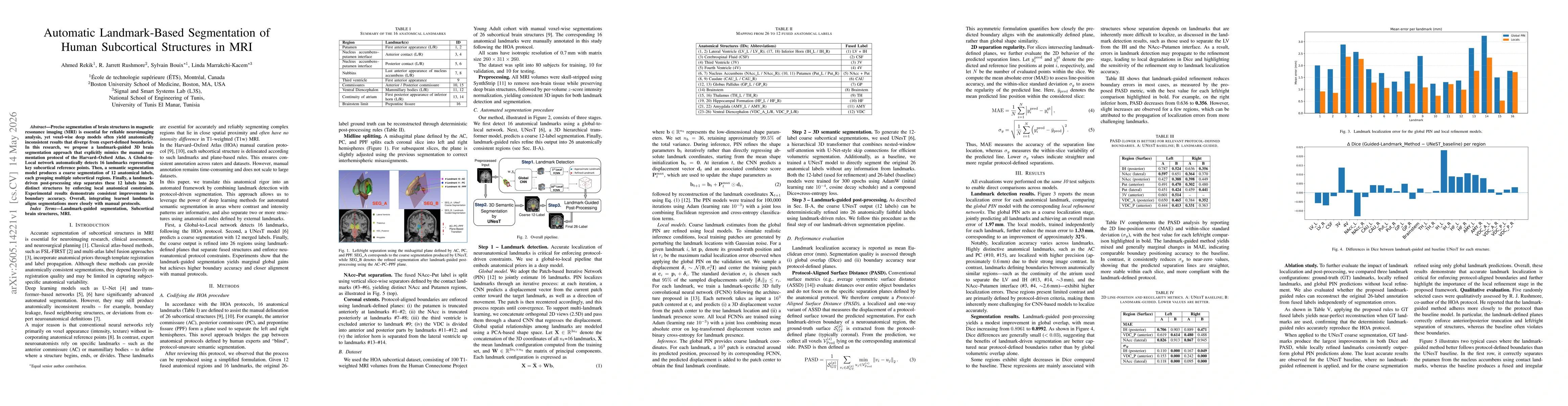
The approach combines a Global-to-Local landmark detection network to locate 16 HOA-defined anatomical landmarks, a UNesT-based 3D hierarchical transformer segmentation model to produce a coarse 12-label segmentation, and a landmark-driven post-processing step that splits the 12 labels into 26 anatomically distinct structures by enforcing plane-based, landmark-defined constraints aligned with the HOA protocol.
Discussion 0