Summary
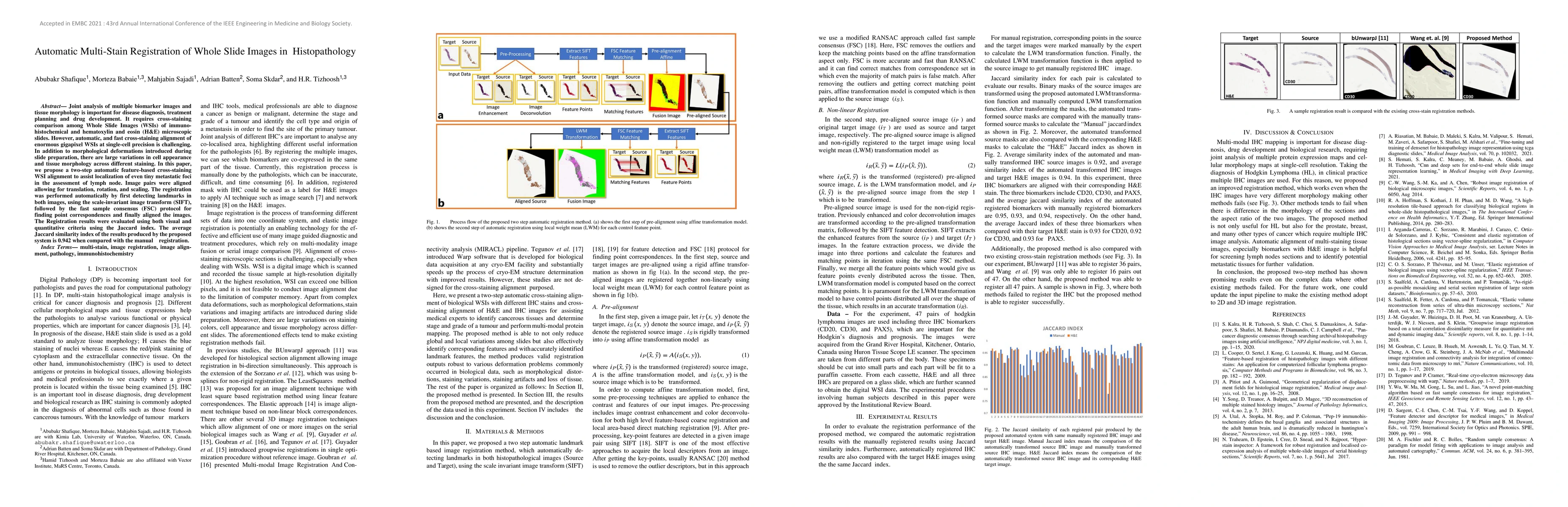
Joint analysis of multiple biomarker images and tissue morphology is important for disease diagnosis, treatment planning and drug development. It requires cross-staining comparison among Whole Slide Images (WSIs) of immuno-histochemical and hematoxylin and eosin (H&E) microscopic slides. However, automatic, and fast cross-staining alignment of enormous gigapixel WSIs at single-cell precision is challenging. In addition to morphological deformations introduced during slide preparation, there are large variations in cell appearance and tissue morphology across different staining. In this paper, we propose a two-step automatic feature-based cross-staining WSI alignment to assist localization of even tiny metastatic foci in the assessment of lymph node. Image pairs were aligned allowing for translation, rotation, and scaling. The registration was performed automatically by first detecting landmarks in both images, using the scale-invariant image transform (SIFT), followed by the fast sample consensus (FSC) protocol for finding point correspondences and finally aligned the images. The Registration results were evaluated using both visual and quantitative criteria using the Jaccard index. The average Jaccard similarity index of the results produced by the proposed system is 0.942 when compared with the manual registration.
AI Key Findings
Get AI-generated insights about this paper's methodology, results, and significance.
Paper Details
PDF Preview
Key Terms
Citation Network
Current paper (gray), citations (green), references (blue)
Display is limited for performance on very large graphs.
Similar Papers
Found 4 papersRegion-guided CycleGANs for Stain Transfer in Whole Slide Images
Stergios Christodoulidis, Maria Vakalopoulou, Eric Deutsch et al.
MUSTANG: Multi-Stain Self-Attention Graph Multiple Instance Learning Pipeline for Histopathology Whole Slide Images
Luca Rossi, Michael Barnes, Gregory Slabaugh et al.
An End-to-end Pipeline for 3D Slide-wise Multi-stain Renal Pathology Registration
Yuankai Huo, Ruining Deng, Peize Li
| Title | Authors | Year | Actions |
|---|
Comments (0)