Purpose: Accurate intraoperative X-ray/CT registration is essential for
surgical navigation in orthopedic procedures. However, existing methods
struggle with consistently achieving sub-millimeter accuracy, robustness under
broad initial pose estimates or need manual key-point annotations. This work
aims to address these challenges by proposing a novel multi-view X-ray/CT
registration method for intraoperative bone registration. Methods: The proposed
registration method consists of a multi-view, contour-based iterative closest
point (ICP) optimization. Unlike previous methods, which attempt to match bone
contours across the entire silhouette in both imaging modalities, we focus on
matching specific subcategories of contours corresponding to bone
substructures. This leads to reduced ambiguity in the ICP matches, resulting in
a more robust and accurate registration solution. This approach requires only
two X-ray images and operates fully automatically. Additionally, we contribute
a dataset of 5 cadaveric specimens, including real X-ray images, X-ray image
poses and the corresponding CT scans. Results: The proposed registration method
is evaluated on real X-ray images using mean reprojection error (mRPD). The
method consistently achieves sub-millimeter accuracy with a mRPD 0.67mm
compared to 5.35mm by a commercial solution requiring manual intervention.
Furthermore, the method offers improved practical applicability, being fully
automatic. Conclusion: Our method offers a practical, accurate, and efficient
solution for multi-view X-ray/CT registration in orthopedic surgeries, which
can be easily combined with tracking systems. By improving registration
accuracy and minimizing manual intervention, it enhances intraoperative
navigation, contributing to more accurate and effective surgical outcomes in
computer-assisted surgery (CAS).
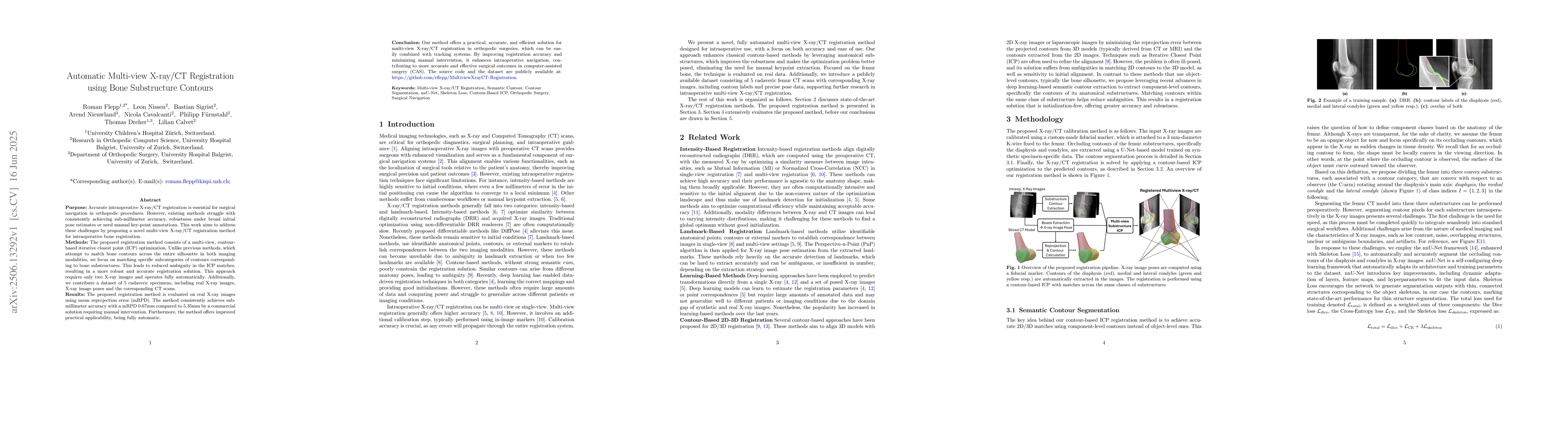
Discussion 0