Authors
Summary
Fetal gestational age (GA) is vital clinical information that is estimated during pregnancy in order to assess fetal growth. This is usually performed by measuring the crown-rump-length (CRL) on an ultrasound image in the Dating scan which is then correlated with fetal age and growth trajectory. A major issue when performing the CRL measurement is ensuring that the image is acquired at the correct view, otherwise it could be misleading. Although clinical guidelines specify the criteria for the correct CRL view, sonographers may not regularly adhere to such rules. In this paper, we propose a new deep learning-based solution that is able to verify the adherence of a CRL image to clinical guidelines in order to assess image quality and facilitate accurate estimation of GA. We first segment out important fetal structures then use the localized structures to perform a clinically-guided mapping that verifies the adherence of criteria. The segmentation method combines the benefits of Convolutional Neural Network (CNN) and the Vision Transformer (ViT) to segment fetal structures in ultrasound images and localize important fetal landmarks. For segmentation purposes, we compare our proposed work with UNet and show that our CNN/ViT-based method outperforms an optimized version of UNet. Furthermore, we compare the output of the mapping with classification CNNs when assessing the clinical criteria and the overall acceptability of CRL images. We show that the proposed mapping is not only explainable but also more accurate than the best performing classification CNNs.
AI Key Findings
Generated Jun 11, 2025
Methodology
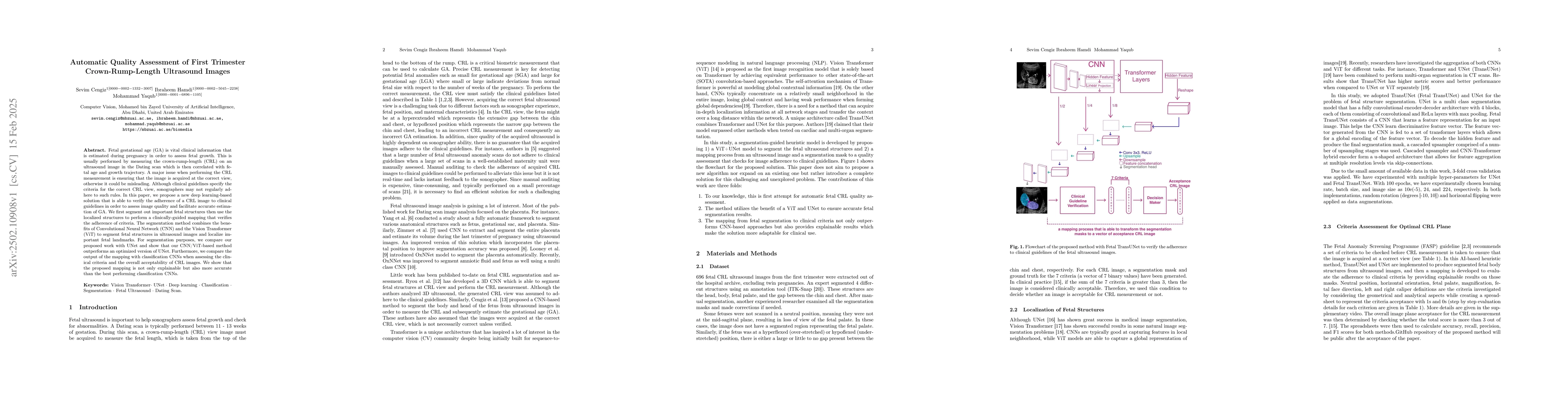
The research proposes a deep learning-based solution for assessing the quality of first trimester crown-rump-length ultrasound images by segmenting fetal structures using a CNN/ViT hybrid method and then verifying adherence to clinical guidelines via a clinically-guided mapping.
Key Results
- A CNN/ViT-based segmentation method outperforms an optimized UNet for fetal structure segmentation in ultrasound images.
- The proposed clinically-guided mapping for assessing CRL image quality is more accurate and explainable than classification CNNs.
- The method facilitates accurate estimation of gestational age by ensuring images adhere to clinical guidelines.
Significance
This research is important for improving the accuracy of gestational age estimation by ensuring correct CRL image acquisition, which can impact fetal growth assessments and pregnancy management.
Technical Contribution
The hybrid CNN/ViT segmentation method combined with a clinically-guided mapping for assessing CRL ultrasound image quality.
Novelty
The proposed approach combines the strengths of CNNs and ViTs for segmentation, outperforming traditional methods like UNet, and introduces an explainable, accurate mapping for clinical guideline adherence assessment.
Limitations
- The paper does not discuss potential limitations of the method in real-world, diverse ultrasound datasets.
- No information is provided on the generalizability of the model to various ultrasound equipment and image qualities.
Future Work
- Investigate the model's performance on diverse ultrasound datasets from different equipment and imaging conditions.
- Explore integration of the method into clinical workflows for real-time quality assessment during ultrasound examinations.
Paper Details
PDF Preview
Citation Network
Current paper (gray), citations (green), references (blue)
Display is limited for performance on very large graphs.
Similar Papers
Found 4 papersDeep Learning-based Quality Assessment of Clinical Protocol Adherence in Fetal Ultrasound Dating Scans
Mohammad Yaqub, Sevim Cengiz
2nd trimester ultrasound (anomaly).
Carocha, Ana, Vicente, Maria, Bernardeco, Joana et al.
Ultrasound-QBench: Can LLMs Aid in Quality Assessment of Ultrasound Imaging?
Zhi Liu, Jun Jia, Guangtao Zhai et al.
| Title | Authors | Year | Actions |
|---|
Comments (0)