Automatic Quantitative Analysis of Brain Organoids via Deep Learning
Publication
Metrics
AI Quick Summary
This paper proposes an automated deep learning method for quantitative analysis of brain organoids, aiming to streamline the observation and analysis of their internal structures. The method was applied to microscopy images of two channels, showing significant differences between Wild Type and Mutant Type organoids.
Paper Preview
Abstract
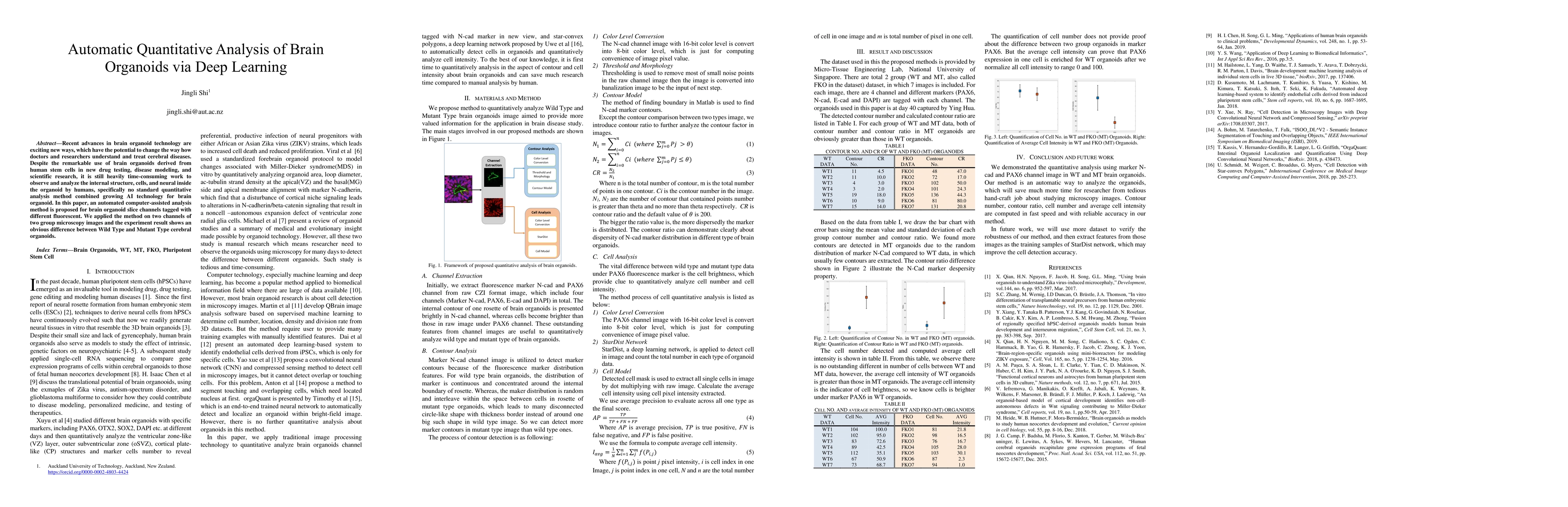
Recent advances in brain organoid technology are exciting new ways, which have the potential to change the way how doctors and researchers understand and treat cerebral diseases. Despite the remarkable use of brain organoids derived from human stem cells in new drug testing, disease modeling, and scientific research, it is still heavily time-consuming work to observe and analyze the internal structure, cells, and neural inside the organoid by humans, specifically no standard quantitative analysis method combined growing AI technology for brain organoid. In this paper, an automated computer-assisted analysis method is proposed for brain organoid slice channels tagged with different fluorescent. We applied the method on two channels of two group microscopy images and the experiment result shows an obvious difference between Wild Type and Mutant Type cerebral organoids.
AI Key Findings
Get AI-generated insights about this paper's methodology, results, significance, and more — seven facets brought into focus.
Impact
Paper Details
Authors
PDF Preview
Key Terms
Citation Network
Current paper (gray), citations (green), references (blue)
Display is limited for performance on very large graphs.
Discussion 0