Automatic Segmentation of Choroid Layer in EDI OCT Images Using Graph Theory in Neutrosophic Space
Publication
Metrics
AI Quick Summary
This paper proposes an automatic method for segmenting the choroid layer in Enhanced Depth Imaging Optical Coherence Tomography (EDI OCT) images using graph theory in neutrosophic space. The method localizes the Retinal Pigment Epithelium layer, applies gamma correction and homomorphic filtering, and demonstrates superior accuracy compared to manual segmentation and other methods.
Paper Preview
Abstract
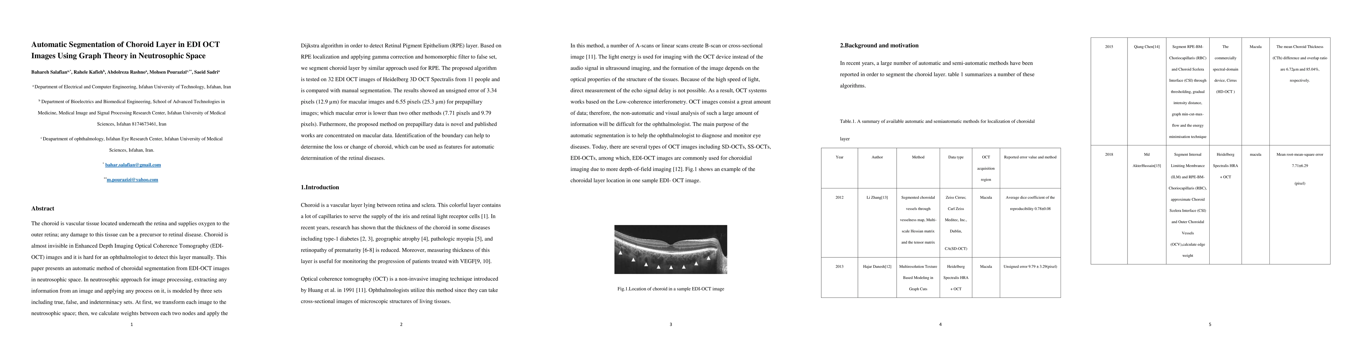
The choroid is vascular tissue located underneath the retina and supplies oxygen to the outer retina; any damage to this tissue can be a precursor to retinal disease. Choroid is almost invisible in Enhanced Depth Imaging Optical Coherence Tomography (EDI-OCT) images and it is hard for an ophthalmologist to detect this layer manually. This paper presents an automatic method of choroidal segmentation from EDI-OCT images in neutrosophic space. In neutrosophic approach for image processing, extracting any information from an image and applying any process on it, is modeled by three sets including true, false, and indeterminacy sets. At first, we transform each image to the neutrosophic space; then, we calculate weights between each two nodes and apply the Dijkstra algorithm in order to detect Retinal Pigment Epithelium (RPE) layer. Based on RPE localization and applying gamma correction and homomorphic filter to false set, we segment choroid layer by similar approach used for RPE. The proposed algorithm is tested on 32 EDI OCT images of Heidelberg 3D OCT Spectralis from 11 people and is compared with manual segmentation. The results showed an unsigned error of 3.34 pixels (12.9 Micrometer) for macular images and 6.55 pixels (25.3 Micrometer) for prepapillary images; which macular error is lower than two other methods (7.71 pixels and 9.79 pixels). Furthermore, the proposed method on prepapillary data is novel and published works are concentrated on macular data. Identification of the boundary can help to determine the loss or change of choroid, which can be used as features for automatic determination of the retinal diseases.
AI Key Findings
Get AI-generated insights about this paper's methodology, results, significance, and more — seven facets brought into focus.
Impact
Paper Details
PDF Preview
Key Terms
Citation Network
Current paper (gray), citations (green), references (blue)
Display is limited for performance on very large graphs.
Discussion 0