Summary
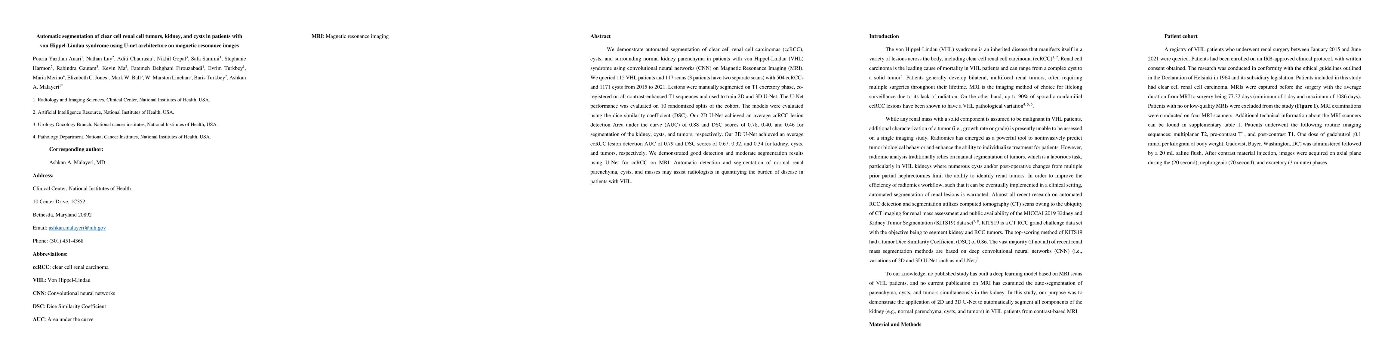
We demonstrate automated segmentation of clear cell renal cell carcinomas (ccRCC), cysts, and surrounding normal kidney parenchyma in patients with von Hippel-Lindau (VHL) syndrome using convolutional neural networks (CNN) on Magnetic Resonance Imaging (MRI). We queried 115 VHL patients and 117 scans (3 patients have two separate scans) with 504 ccRCCs and 1171 cysts from 2015 to 2021. Lesions were manually segmented on T1 excretory phase, co-registered on all contrast-enhanced T1 sequences and used to train 2D and 3D U-Net. The U-Net performance was evaluated on 10 randomized splits of the cohort. The models were evaluated using the dice similarity coefficient (DSC). Our 2D U-Net achieved an average ccRCC lesion detection Area under the curve (AUC) of 0.88 and DSC scores of 0.78, 0.40, and 0.46 for segmentation of the kidney, cysts, and tumors, respectively. Our 3D U-Net achieved an average ccRCC lesion detection AUC of 0.79 and DSC scores of 0.67, 0.32, and 0.34 for kidney, cysts, and tumors, respectively. We demonstrated good detection and moderate segmentation results using U-Net for ccRCC on MRI. Automatic detection and segmentation of normal renal parenchyma, cysts, and masses may assist radiologists in quantifying the burden of disease in patients with VHL.
AI Key Findings
Get AI-generated insights about this paper's methodology, results, and significance.
Paper Details
PDF Preview
Key Terms
Citation Network
Current paper (gray), citations (green), references (blue)
Display is limited for performance on very large graphs.
Similar Papers
Found 4 papersThe gradual expansion of multiple intramedullary metastatic renal cell carcinoma in a patient with von Hippel-Lindau disease with multiple intramedullary hemangioblastomas: illustrative case.
Miki, Shunichiro, Sakamoto, Noriaki, Matsuda, Masahide et al.
Characterization of complex renal cysts in hereditary leiomyomatosis and renal cell cancerUsing magnetic resonance based qualitative features.
Linehan, W Marston, Ball, Mark W, Sheikhy, Ali et al.
| Title | Authors | Year | Actions |
|---|
Comments (0)