Automatic Segmentation of the Great Arteries for Computational Hemodynamic Assessment
Publication
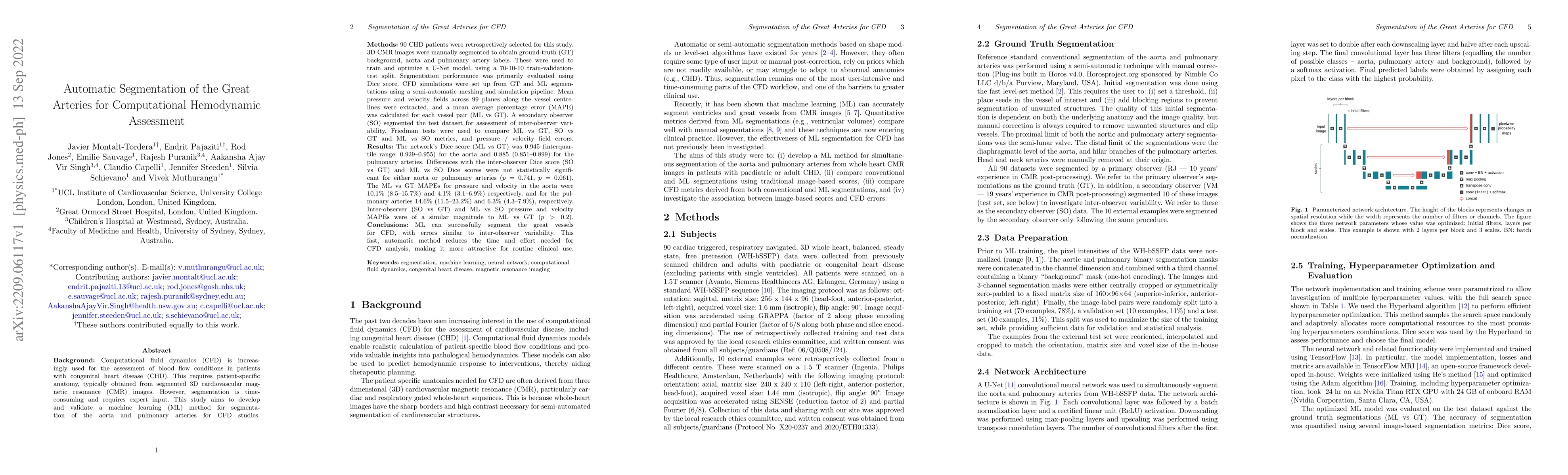
Metrics
AI Quick Summary
This study developed a machine learning method to automatically segment the aorta and pulmonary arteries for computational fluid dynamics in congenital heart disease patients, using 3D cardiovascular magnetic resonance images. The U-Net model achieved high segmentation accuracy comparable to inter-observer variability, reducing the need for manual segmentation and thus the time required for computational hemodynamic assessments.
Paper Preview
Abstract
Background: Computational fluid dynamics (CFD) is increasingly used to assess blood flow conditions in patients with congenital heart disease (CHD). This requires patient-specific anatomy, usually obtained from segmented 3D cardiovascular magnetic resonance (CMR) images. However, segmentation is time-consuming and needs expert input. This study aims to develop and validate a machine learning (ML) method for segmentation of the aorta and pulmonary arteries (PAs) for CFD studies. Methods: 90 CHD patients were retrospectively selected for this study. 3D CMR images were manually segmented to obtain ground-truth (GT) background, aorta and PA labels. These were used to train and optimize a U-Net model. Segmentation performance was primarily evaluated using Dice score. CFD simulations were set up from GT and ML segmentations using a semi-automatic meshing and simulation pipeline. Pressure and velocity fields were computed, and a mean average percentage error (MAPE) was calculated for each vessel pair. A secondary observer (SO) segmented the test dataset to assess inter-observer variability. Friedman tests were used to compare segmentation metrics and flow field errors. Results: The model's Dice score (ML vs GT) was 0.945 for the aorta and 0.885 for the PAs. Differences with the inter-observer Dice score (SO vs GT) and ML vs SO Dice scores were not statistically significant for either aorta or PAs. The ML vs GT MAPEs for pressure and velocity in the aorta were 10.1% and 4.1% respectively, and for the PAs 14.6% and 6.3%, respectively. Inter-observer (SO vs GT) and ML vs SO pressure and velocity MAPEs were of a similar magnitude to ML vs GT. Conclusions: The proposed method can successfully segment the great vessels for CFD, with errors similar to inter-observer variability. This reduces the time and effort needed for CFD analysis, making it more attractive for routine clinical use.
AI Key Findings
Get AI-generated insights about this paper's methodology, results, significance, and more — seven facets brought into focus.
Impact
Paper Details
Authors
PDF Preview
Key Terms
Citation Network
Current paper (gray), citations (green), references (blue)
Display is limited for performance on very large graphs.
Discussion 0