Automatic Segmentation of the Left Ventricle in Cardiac CT Angiography Using Convolutional Neural Network
Publication
Metrics
AI Quick Summary
This paper presents an automatic method for segmenting the left ventricle in cardiac CT angiography using convolutional neural networks, achieving an average Dice coefficient of 0.85 and a mean absolute surface distance of 1.1 mm, demonstrating the feasibility of the approach.
Paper Preview
Abstract
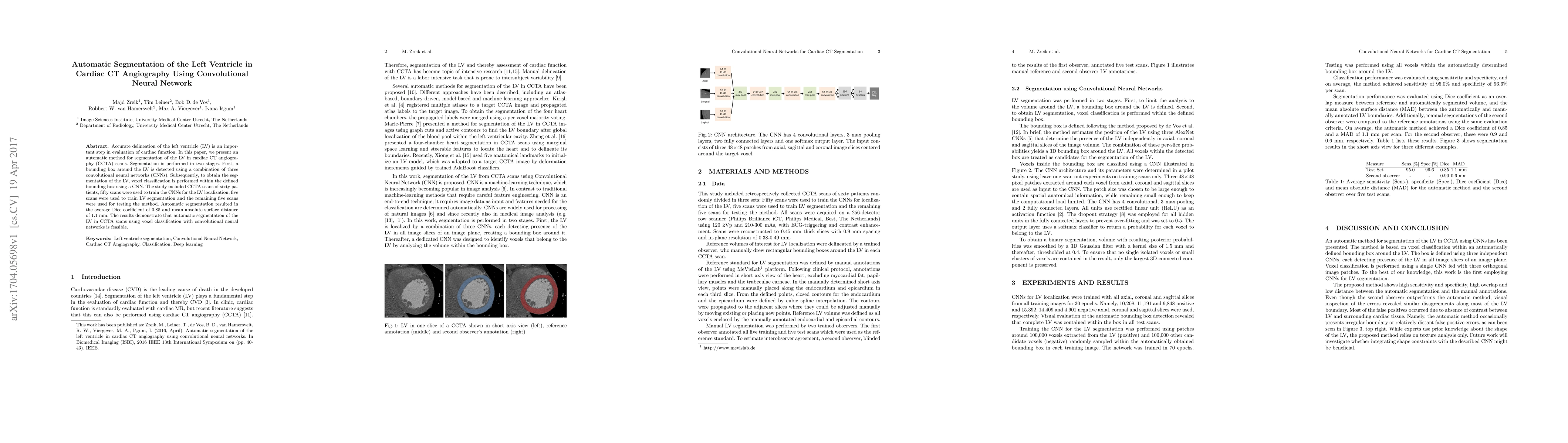
Accurate delineation of the left ventricle (LV) is an important step in evaluation of cardiac function. In this paper, we present an automatic method for segmentation of the LV in cardiac CT angiography (CCTA) scans. Segmentation is performed in two stages. First, a bounding box around the LV is detected using a combination of three convolutional neural networks (CNNs). Subsequently, to obtain the segmentation of the LV, voxel classification is performed within the defined bounding box using a CNN. The study included CCTA scans of sixty patients, fifty scans were used to train the CNNs for the LV localization, five scans were used to train LV segmentation and the remaining five scans were used for testing the method. Automatic segmentation resulted in the average Dice coefficient of 0.85 and mean absolute surface distance of 1.1 mm. The results demonstrate that automatic segmentation of the LV in CCTA scans using voxel classification with convolutional neural networks is feasible.
AI Key Findings
Get AI-generated insights about this paper's methodology, results, significance, and more — seven facets brought into focus.
Impact
Paper Details
PDF Preview
Key Terms
Citation Network
Current paper (gray), citations (green), references (blue)
Display is limited for performance on very large graphs.
Discussion 0