ArxivLens

Publication

Published: Jan 20, 2026
Categories: physics.med-ph

Metrics

Source: ArXiv

Quick Answers

What methodology did the authors use?

The study employs a combination of advanced computational techniques including full-waveform inversion (FWI), machine learning, and physics-based modeling to reconstruct images from ultrasound data. It integrates SE-Net architectures with FWI to enhance image resolution and accuracy. More in Methodology →

What are the key results?

Successful reconstruction of high-resolution images from sparse ultrasound data using the proposed method — Significant improvement in image quality compared to traditional FWI methods More in Key Results →

Why is this work significant?

This research advances medical imaging by providing a more accurate and efficient method for reconstructing images from limited ultrasound data, which could improve diagnostic capabilities and reduce scanning times in clinical settings. More in Significance →

What are the main limitations?

The method requires substantial computational resources which may limit its practical implementation in resource-constrained environments — Validation was primarily based on simulated data rather than extensive clinical trials More in Limitations →

Paper Preview

Abstract

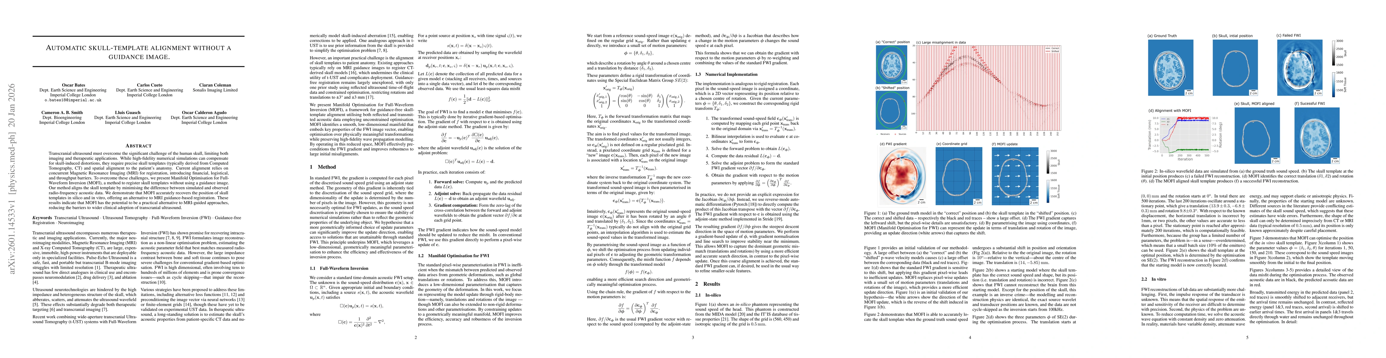
Transcranial ultrasound must overcome the significant challenge of the human skull, limiting both imaging and therapeutic applications. While high-fidelity numerical simulations can compensate for skull-induced distortions, they require precise skull templates (typically derived from Computed Tomography, CT) and spatial alignment to the patient's anatomy. Current alignment relies on concurrent Magnetic Resonance Imaging (MRI) for registration, introducing financial, logistical, and throughput barriers. To overcome these challenges, we present Manifold Optimisation for Full-Waveform Inversion (MOFI), a method to register skull templates without using a guidance image. Our method aligns the skull template by minimising the difference between simulated and observed radio-frequency acoustic data. We demonstrate that MOFI accurately recovers the position of skull templates in silico and in vitro, offering an alternative to MRI guidance-based registration. These results indicate that MOFI has the potential to be a practical alternative to MRI-guided approaches, reducing the barriers to wider clinical adoption of transcranial ultrasound.

Key Findings, in focus

Seven facets of this paper, analysed and brought into focus by AI.

AI-generated · verify against source Generated Jan 22, 2026
In focus · one-line synthesis

This research advances medical imaging by providing a more accurate and efficient method for reconstructing images from limited ultrasound data, which could improve diagnostic capabilities and reduce scanning times in clinical settings.

01
MethodologyHow they did it

The study employs a combination of advanced computational techniques including full-waveform inversion (FWI), machine learning, and physics-based modeling to reconstruct images from ultrasound data. It integrates SE-Net architectures with FWI to enhance image resolution and accuracy.

02
ResultsWhat they found
  • Successful reconstruction of high-resolution images from sparse ultrasound data using the proposed method
  • Significant improvement in image quality compared to traditional FWI methods
  • Demonstration of the method's effectiveness in transcranial ultrasound imaging applications
03
SignificanceWhy it matters

This research advances medical imaging by providing a more accurate and efficient method for reconstructing images from limited ultrasound data, which could improve diagnostic capabilities and reduce scanning times in clinical settings.

04
Technical ContributionsWhat's new technically

The integration of SE-Net architectures with full-waveform inversion represents a novel technical contribution, enabling more accurate and efficient image reconstruction from sparse ultrasound data through enhanced feature extraction and learning capabilities.

05
NoveltyWhat sets it apart

This work introduces a novel hybrid approach combining deep learning with physics-based modeling, which differs from traditional methods that rely solely on either data-driven or physics-based approaches.

06
LimitationsWhat to be cautious about
  • The method requires substantial computational resources which may limit its practical implementation in resource-constrained environments
  • Validation was primarily based on simulated data rather than extensive clinical trials
07
Future WorkWhere it goes next
  • Exploring the application of this methodology in other medical imaging modalities
  • Developing more efficient computational frameworks to reduce resource requirements
  • Conducting large-scale clinical trials to validate the method's effectiveness in real-world scenarios

Paper Details

Paper ID: 2601.14533
Categories:
physics.med-ph

How to Cite This Paper

Reference manager: RIS EndNote


Discussion 0

This website uses cookies to ensure you get the best experience. Learn more