Automatic Tumor Segmentation via False Positive Reduction Network for Whole-Body Multi-Modal PET/CT Images
Publication
Metrics
AI Quick Summary
This paper proposes a deep learning method for automatic tumor segmentation in whole-body PET/CT images, focusing on reducing false positives. The method uses a self-supervised pre-trained global segmentation module followed by a local refinement module to accurately delineate tumor regions, achieving a top-ranked dice score of 0.9324 in preliminary tests.
Paper Preview
Abstract
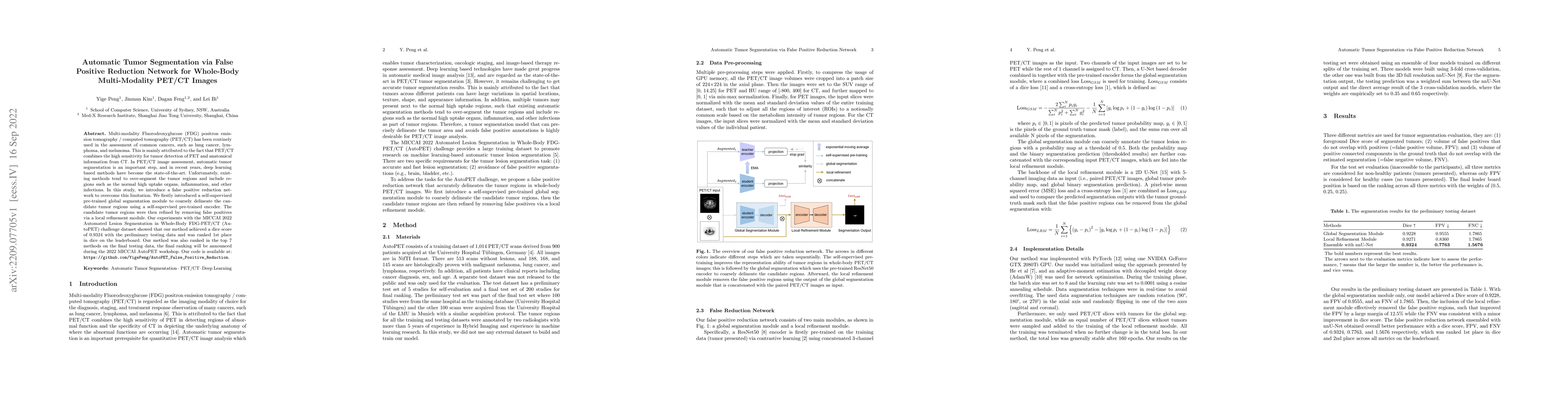
Multi-modality Fluorodeoxyglucose (FDG) positron emission tomography / computed tomography (PET/CT) has been routinely used in the assessment of common cancers, such as lung cancer, lymphoma, and melanoma. This is mainly attributed to the fact that PET/CT combines the high sensitivity for tumor detection of PET and anatomical information from CT. In PET/CT image assessment, automatic tumor segmentation is an important step, and in recent years, deep learning based methods have become the state-of-the-art. Unfortunately, existing methods tend to over-segment the tumor regions and include regions such as the normal high uptake organs, inflammation, and other infections. In this study, we introduce a false positive reduction network to overcome this limitation. We firstly introduced a self-supervised pre-trained global segmentation module to coarsely delineate the candidate tumor regions using a self-supervised pre-trained encoder. The candidate tumor regions were then refined by removing false positives via a local refinement module. Our experiments with the MICCAI 2022 Automated Lesion Segmentation in Whole-Body FDG-PET/CT (AutoPET) challenge dataset showed that our method achieved a dice score of 0.9324 with the preliminary testing data and was ranked 1st place in dice on the leaderboard. Our method was also ranked in the top 7 methods on the final testing data, the final ranking will be announced during the 2022 MICCAI AutoPET workshop. Our code is available at: https://github.com/YigePeng/AutoPET_False_Positive_Reduction.
AI Key Findings
Get AI-generated insights about this paper's methodology, results, significance, and more — seven facets brought into focus.
Impact
Paper Details
Authors
PDF Preview
Key Terms
Citation Network
Current paper (gray), citations (green), references (blue)
Display is limited for performance on very large graphs.
Discussion 0